北京大学学报(医学版) ›› 2019, Vol. 51 ›› Issue (1): 59-64. doi: 10.19723/j.issn.1671-167X.2019.01.011
近红外光透照技术诊断乳磨牙早期邻面龋
周琼,彭楚芳,秦满( )
)
- 北京大学口腔医学院·口腔医院,儿童口腔科 国家口腔疾病临床医学研究中心 口腔数字化医疗技术和材料国家工程实验室 口腔数字医学北京市重点实验室, 北京 100081
Near-infrared light transillumination for detection of incipient proximal caries in primary molars
Qiong ZHOU,Chu-fang PENG,Man QIN()
- Department of Pediatric Dentistry, Peking University School and Hospital of Stomatology & National Clinical Research Center for Oral Diseases & National Engineering Laboratory for Digital and Material Technology of Stomatology & Beijing Key Laboratory of Digital Stomatology, Beijing 100081, China
摘要:
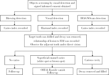
目的:评价近红外光透照技术(near-infrared light transillumination,NILT,商品名DIAGNOcam)诊断乳磨牙早期邻面龋的应用价值。方法:对北京大学口腔医院儿童口腔科4~9岁疑有乳磨牙早期邻面龋的患儿进行视诊、咬合翼片和DIAGNOcam检查,龋齿指数记录为0(无龋)、1(釉质浅层龋)、2(釉质深层龋不超出釉牙本质界)、3(牙本质龋)。相邻2颗乳磨牙中至少1颗牙齿符合侵入性治疗标准方可纳入研究,即要求至少两项检查的龋齿指数≥2。对龋齿指数≥2的牙齿行侵入性治疗,直视下观察相邻乳磨牙的情况,若龋坏成洞,则也行侵入性治疗。将直视下龋洞深度与釉牙本质界的关系作为金标准,比较3种检查方法诊断乳磨牙早期邻面龋的灵敏度、特异度和准确度。结果:共纳入36例患儿104个乳磨牙邻面,其中,5个牙面最终诊断为无龋,20个牙面诊断为釉质浅层龋,79个牙面进行了侵入性治疗,包括22个牙面龋洞达釉牙本质界,57个牙面龋洞达牙本质层内。视诊、咬合翼片和DIAGNOcam诊断乳磨牙早期邻面龋的灵敏度为68.69%、81.82%、78.79%,特异度为100.00%、100.00%、80.00%,准确度为75.96%、85.65%、78.85%。对于釉质浅层龋,视诊诊断的灵敏度最低,仅为10.00%,DIAGNOcam的灵敏度(60.00%)高于咬合翼片(40.00%);对于釉质深层龋的诊断灵敏度,咬合翼片(86.36%)>DIAGNOcam(72.73%)>视诊(68.18%);对于牙本质龋,3种方法的诊断灵敏度均超过85%,咬合翼片最高为94.74%。结论:DIAGNOcam诊断乳磨牙早期邻面龋具有接近于咬合翼片的准确度,且使用安全、简便、无辐射,适用于儿童口腔临床。
中图分类号:
- R781.1




| [1] | 葛立宏 . 儿童口腔医学 [M]. 北京: 人民卫生出版社, 2012: 113. |
| [2] |
Verdonschot EH, Angmar-Månsson B, ten Bosch JJ , et al. Deve-lopments in caries diagnosis and their relationship to treatment decisions and quality of care. ORCA Saturday Afternoon Symposium 1997[J]. Caries Res, 1999,33(1):32-40.
doi: 10.1159/000016493 |
| [3] |
Wenzel A . Bitewing and digital bitewing radiography for detection of caries lesions[J]. J Dent Res, 2004,83:C72-C75.
doi: 10.1177/154405910408301S14 pmid: 15286126 |
| [4] |
Friedman J, Marcus MI . Transillumination of theoral cavity with use of fiber optics[J]. J Am Dent Assoc, 1970,80(4):801-809.
doi: 10.14219/jada.archive.1970.0117 pmid: 5264574 |
| [5] | 陈君, 岳林 . 对光纤透照法诊断后牙邻面龋的评价[J]. 现代口腔医学杂志, 1998,12(3):178-180. |
| [6] |
Ye SY, Kim JY, Ro JH , et al. Detecting incipient caries using front-illuminated infrared light scattering imaging[J]. Trans Electr Electron Mater, 2012,13(6):310-316.
doi: 10.4313/TEEM.2012.13.6.310 |
| [7] |
Astvaldsdóttir A, Ahlund K, Holbrook WP , et al. Approximal caries detection by DIFOTI: in vitro comparison of diagnostic accuracy/efficacy with film and digital radiography[J]. Int J Dent, 2012,326401. doi: 10.1155/2012/326401.
doi: 10.1155/2012/326401 pmid: 23213335 |
| [8] |
Abdelaziz M, Krejci I . DIAGNOcam—a near infrared digital imaging transillumination (NIDIT) technology[J]. Int J Esthet Dent, 2015,10(1):158-165.
pmid: 25625132 |
| [9] |
高洁, 李波, 刘月华 . 乳切牙牙体硬组织的扫描电镜观察及临床分析[J]. 实用口腔医学杂志, 2008,24(2):274-276.
doi: 10.3969/j.issn.1001-3733.2008.02.029 |
| [10] | 于世凤 . 口腔组织病理学 [M]. 北京: 人民卫生出版社, 2006: 141-147. |
| [11] |
Abogazalah N, Eckert GJ, Ando M . In vitro performance of near infrared light transillumination at 780-nm and digital radiography for detection of non-cavitated approximal caries[J]. J Dent, 2017,63:44-50.
doi: 10.1016/j.jdent.2017.05.018 pmid: 28559050 |
| [12] |
Kühnisch J, Söchtig F, Pitchika V , et. al. In vivo validation of near-infrared light transillumination for interproximal dentin caries detection[J]. Clin Oral Investig, 2016,20(4):821-829.
doi: 10.1007/s00784-015-1559-4 pmid: 26374746 |
| [13] |
Mejàre I, Gröndahl HG, Carlstedt K , et al. Accuracy at radiography and probing for the diagnosis of proximal caries[J]. Scand J Dent Res, 1985,93(2):178-184.
doi: 10.1111/j.1600-0722.1985.tb01328.x pmid: 3858967 |
| [14] |
于江利, 唐仁韬, 冯琳 , 等. 数字化光纤透照法判断龋洞深度[J]. 北京大学学报(医学版), 2017,49(1):81-85.
doi: 10.3969/j.issn.1671-167X.2017.01.014 |
| [1] | 马豆豆, 马晓彩, 常天静, 王丽芳, 丁艳, 石连杰. 临床表现似系统性红斑狼疮的大B细胞淋巴瘤骨髓受累1例[J]. 北京大学学报(医学版), 2026, 58(3): 666-669. |
| [2] | 赵业, 刁小莉, 熊焰. 细胞转移技术在微量细胞液病理诊断中的应用[J]. 北京大学学报(医学版), 2026, 58(1): 208-213. |
| [3] | 王月, 梁宇红. 繁茂型牙骨质-骨结构不良1例[J]. 北京大学学报(医学版), 2026, 58(1): 220-224. |
| [4] | 池彦廷, 蒋鸿杰, 陈艳, 徐志秀, 李斌斌. 直接免疫荧光在口腔黏膜寻常型天疱疮诊断中的价值: 基于多指标联合分析的回顾性研究[J]. 北京大学学报(医学版), 2026, 58(1): 68-73. |
| [5] | 顾静妍, 李欣艺, 赵金霞, 穆荣. 误诊为类风湿关节炎、痛风的糖尿病致Charcot关节病1例[J]. 北京大学学报(医学版), 2025, 57(6): 1193-1197. |
| [6] | 肖晓笛, 夏有辰, 柳剑英, 付鹏. 左侧胸锁乳突肌间血管内乳头状内皮增生1例[J]. 北京大学学报(医学版), 2025, 57(5): 1002-1004. |
| [7] | 孙翔宇, 袁超, 周芯竹, 刁婧, 郑树国. 唾液微生态在口腔及全身疾病早期防治中的应用[J]. 北京大学学报(医学版), 2025, 57(5): 859-863. |
| [8] | 马丽娟, 腾雍辉, 王勇, 赵一姣, 张馨月, 秦庆钊, 尹东. 乳牙缺失数字化丝圈间隙保持器的三维有限元分析[J]. 北京大学学报(医学版), 2025, 57(2): 376-383. |
| [9] | 陈钊, 邱永康, 康磊. 经典型Sweet综合征 18F-FDG PET/CT多脏器异常显像1例[J]. 北京大学学报(医学版), 2025, 57(2): 403-407. |
| [10] | 方媛媛, 徐帆, 雷杰, 张昊, 张文宇, 孙宇, 吴宏新, 傅开元, 毛伟玉. 基于颞下颌关节紊乱病诊断标准的临床自动诊断系统的建立及验证[J]. 北京大学学报(医学版), 2025, 57(1): 192-201. |
| [11] | 车佳璐, 刘子臣, 李琨, 张晨, 车南颖. 全自动EasyNAT核酸快速检测系统检测石蜡包埋组织诊断结核病的临床价值[J]. 北京大学学报(医学版), 2024, 56(6): 1047-1051. |
| [12] | 陈心心, 唐哲, 乔艳春, 荣文笙. 北京市密云区4岁儿童患龋状况及其与龋活跃性检测的相关性[J]. 北京大学学报(医学版), 2024, 56(5): 833-838. |
| [13] | 钟华, 李原, 徐丽玲, 白明欣, 苏茵. 18F-FDG PET/CT在风湿免疫病中的应用[J]. 北京大学学报(医学版), 2024, 56(5): 853-859. |
| [14] | 李正芳,罗采南,武丽君,吴雪,孟新艳,陈晓梅,石亚妹,钟岩. 抗氨基甲酰化蛋白抗体在诊断类风湿关节炎中的应用价值[J]. 北京大学学报(医学版), 2024, 56(4): 729-734. |
| [15] | 姚海红,杨帆,唐素玫,张霞,何菁,贾园. 系统性红斑狼疮及成人Still病合并巨噬细胞活化综合征的临床特点及诊断指标[J]. 北京大学学报(医学版), 2023, 55(6): 966-974. |
|
||
