北京大学学报(医学版) ›› 2019, Vol. 51 ›› Issue (2): 349-355. doi: 10.19723/j.issn.1671-167X.2019.02.028
无牙颌印模用个别托盘椅旁计算机辅助设计和三维打印系统建立与临床初步评价
王冠博1,叶红强1,陈虎2,王勇2,孙玉春2,∆( ),周永胜1,∆()
- 1. 北京大学口腔医学院·口腔医院,修复科 国家口腔疾病临床医学研究中心 口腔数字化医疗技术和材料国家工程实验室 口腔数字医学北京市重点实验室, 北京 100081;
2. 北京大学口腔医学院·口腔医院口腔医学数字化研究中心 国家口腔疾病临床医学研究中心 口腔数字化医疗技术和材料国家工程实验室 口腔数字医学北京市重点实验室,卫生部口腔医学计算机应用工程技术研究中心,北京 100081;
Establishment and preliminary clinical evaluation of edentulous custom trays designed and fabricated by chair-side CAD and 3D printing systems
Kuan-paul WANG1,Hong-qiang YE1,Hu CHEN2,Yong WANG2,Yu-chun SUN2,∆(),Yong-sheng ZHOU1,∆()
- 1. Department of Prosthodontics, Peking University School and Hospital of Stomatology & National Clinical Research Center for Oral Diseases & National Engineering Laboratory for Digital and Material Technology of Stomatology & Beijing Key Laboratory of Digital Stomatology, Beijing 100081, China;
2. Center of Digital Dentistry, Peking University School and Hospital of Stomatology & National Clinical Research Center for Oral Diseases & National Engineering Laboratory for Digital and Material Technology of Stomatology & Beijing Key Laboratory of Digital Stomatology,Research Center of Engineering and Technology for Digital Dentistry, Ministry of Health, Beijing 100081, China;
摘要:
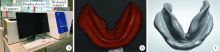
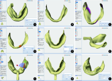
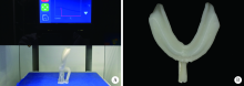
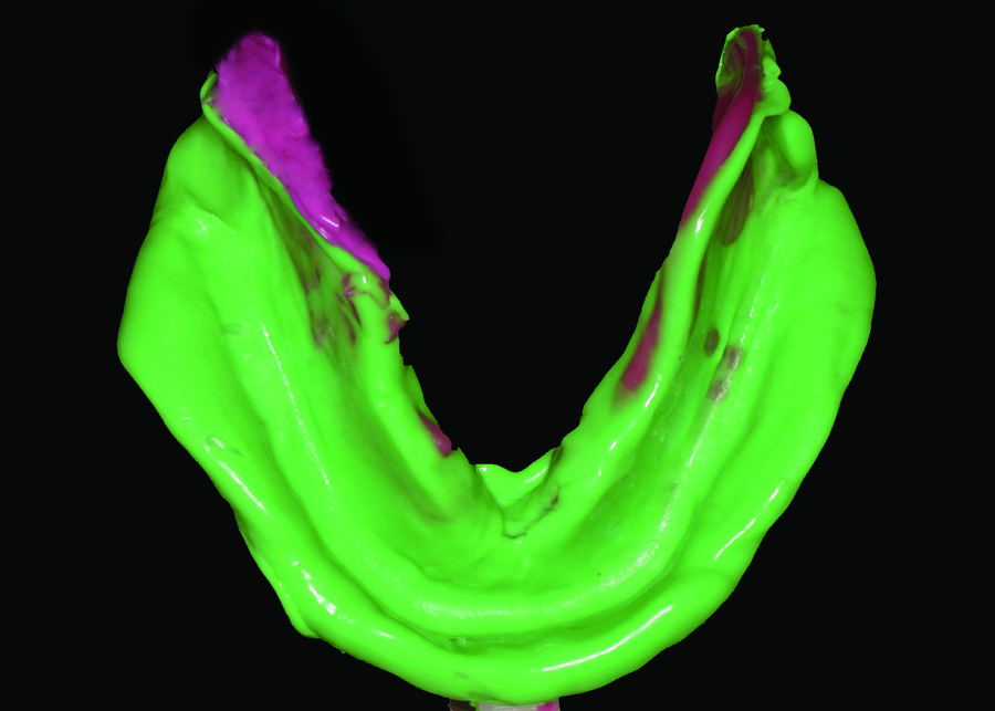
目的: 采用自主研发的无牙颌个别托盘椅旁数字化系统设计并三维打印无牙颌个别托盘,用视觉模拟评分法,基于医师对该个别托盘制取终印模的满意度,初步评价其临床应用效果。方法: 由三位经过统一操作流程培训的修复医师随机接诊于北京大学口腔医院就诊的15名上、下颌牙列缺失患者,分别采用自主研发无牙颌个别托盘椅旁数字化系统和传统手工法制作无牙颌个别托盘,高流动型硅橡胶印模材制取终印模。采用视觉模拟评分法设计调查问卷,由制取终印模的三位医师根据制取印模的满意程度填写调查问卷,进行满意度调查。结果: 根据制取印模的满意程度,使用自主研发无牙颌个别托盘椅旁数字化系统制作的个别托盘与传统手工法制作的无牙颌个别托盘在口腔内试戴(整体尺寸、外形、是否利于握持等)的平均满意度分别为9.18±0.19和8.23±0.22,两者差异有统计学意义(P<0.05);试戴时边缘位置(边缘伸展、系带切迹)的平均满意度分别为8.91±0.40和7.96±0.23,两者差异有统计学意义(P<0.05);托盘稳定性的平均满意度分别为8.80±0.83和8.01±0.81,两者差异有统计学意义(P<0.05);终印模制取效果(完整性、印模材厚度、是否漏出托盘、边缘形态)的平均满意度分别为8.94±0.68和7.99±0.42,两者差异有统计学意义(P<0.05);优质终印模获取难度(重复次数、效率)的平均满意度分别为9.20±0.37和7.88±0.22,两者差异有统计学意义(P<0.05);整体的平均满意度分别为 9.11±0.49和7.95±0.15,两者差异有统计学意义(P<0.05)。结论: 使用自主研发无牙颌个别托盘椅旁数字化系统制作的个别托盘制取终印模的医师满意度高于传统手工光固化法制作的个别托盘, 达到临床要求,可进一步在临床上推广使用。
中图分类号:
- R783.6


| [1] | 冯海兰, 徐军 . 口腔修复学[M]. 2版. 北京: 北京大学医学出版社, 2013: 267-269. |
| [2] | Basker RM, Davenport JC, Thomason JM . Prosthetic treatment of the edentulous patient[M]. 4th ed. Oxford: Blackwell, 2002: 130-149. |
| [3] | Zarb GA, Bolender GL, Eckert SE , et al. Prosthodontic treatment for edentulous patients[M]. 13th ed. St. Louis: Elsevier Mosby, 2013: 170-179. |
| [4] |
Boucher CO . Complete dentureprosthodontics: the state of the art[J]. J Prosthet Dent, 2004,92(4):309-315.
doi: 10.1016/j.prosdent.2004.05.017 |
| [5] | Rahn AO, Ivanhoe JR, Plummer KD. 全口义齿教科书[M]. 6版. 冯海兰,译. 北京: 人民卫生出版社, 2011: 74-79. |
| [6] |
Petrie CS, Walker MP, Williams K . A Survey of U.S. prosthodontists and dental schools on the current materials and methods for final impressions for complete denture prosthodontics[J]. J Prosthodont, 2005,14(4):253-262.
doi: 10.1111/jopr.2005.14.issue-4 |
| [7] | Al-Ahmar AO, Lynch CD, Locke M , et al. Quality of master impressions and related materials for fabrication of complete dentures in the UK[J]. J Oral Rehabil, 2008,35(2):111-115. |
| [8] |
van Noort R . The future of dental devices is digital[J]. Dent Mater, 2012,28(1):3-12.
doi: 10.1016/j.dental.2011.10.014 |
| [9] |
Dawood A, Marti Marti B, Sauret-Jackson V , et al. 3D printing in dentistry[J]. Br Dent J, 2015,219(11):521-529.
doi: 10.1038/sj.bdj.2015.914 |
| [10] | Baroudi K, Ibraheem SN . Assessment of chair-side computer-aided design and computer-aided manufacturing restorations: a review of the literature[J]. J Int Oral Health, 2015,7(4):96-104. |
| [11] | Patzelt SBM, Spies BC, Kohal RJ . CAD/CAM-fabricated implant-supported restorations: a systematic review[J]. Clin Oral Implants Res, 2015,26(Suppl.11):77-85. |
| [12] |
Williams RJ, Bibb R, Eggbeer D , et al. Use of CAD/CAM technology to fabricate a removable partial denture framework[J]. J Prosthet Dent, 2006,96(2):96-99.
doi: 10.1016/j.prosdent.2006.05.029 |
| [13] | 吴琳, 吕培军, 王勇 , 等. 可摘局部义齿支架铸型的计算机辅助设计与制作[J]. 中华口腔医学杂志, 2006,41(7):432-435. |
| [14] | 孙玉春, 吕培军, 王勇 , 等. 计算机辅助设计与快速成形技术辅助制作全口义齿的探讨[J]. 中华口腔医学杂志, 2007,42(6):324-329. |
| [15] |
Chen H, Yang X, Chen L , et al. Application of FDM three-dimensional printing technology in the digital manufacture of custom edentulous mandible trays[J]. Sci Rep, 2016,6:19207.
doi: 10.1038/srep19207 |
| [16] |
陈虎, 赵甜, 王勇 , 等. 基于初印模三维扫描的无牙颌上颌个性化托盘计算机辅助设计及三维打印[J]. 北京大学学报(医学版), 2016,48(5):900-904.
doi: 10.3969/j.issn.1671-167X.2016.05.028 |
| [17] | 魏菱, 陈虎, 周永胜 , 等. 数字化全口义齿个别托盘制作与临床应用时间评价[J]. 北京大学学报(医学版), 2017,49(1):86-91. |
| [18] |
Liu Q, Leu MC, Schmitt SM . Rapid prototyping in dentistry: technology and application[J]. Int J Adv Manuf Technol, 2006,29(3):317-335.
doi: 10.1007/s00170-005-2523-2 |
| [19] | Torabi K, Farjood E, Hamedani S . Rapid prototyping technologies and their applications in prosthodontics, a review of literature[J]. J Dent, 2015,16(1):1-9. |
| [20] | 徐军 . 总义齿与可摘局部义齿的设计[M]. 北京: 中国大百科全书出社, 2005: 16-17. |
| [21] | Chang JJ, Chen JH, Lee HE , et al. Maximizing mandibular denture retention in the sublingual space[J]. Int J Prosthodont, 2011,24(5):460-464. |
| [1] | 白晓强, 袁芷若, 周永胜, 吕珑薇. 动态牵张促进人骨髓间充质干细胞三维培养的成骨分化[J]. 北京大学学报(医学版), 2026, 58(3): 641-649. |
| [2] | 刁畅, 王时敏, 李曼, 潘韶霞, 刘洋. 牙列缺失种植覆盖义齿集中𬌗型的临床研究[J]. 北京大学学报(医学版), 2026, 58(1): 133-138. |
| [3] | 温奥楠, 张晓会, 杨咏涛, 高梓翔, 李文博, 单珅瑶, 商相宜, 田淯文, 郭殊玮, 王艺蓁, 王勇, 赵一姣. 基于非刚性配准构建三维颜面微笑仿真序列数据的方法[J]. 北京大学学报(医学版), 2026, 58(1): 139-144. |
| [4] | 于录, 吴灵, 刘筱菁, 李自力. 基于数据库相似性检索的正颌外科手术规划技术流程可行性研究: 随机对照试验[J]. 北京大学学报(医学版), 2026, 58(1): 145-152. |
| [5] | 邵梁, 马雯洁, 陈莹, 丁茜, 张磊. 上颌切牙前伸和正中咬合接触解剖特征的数字化测量与分析[J]. 北京大学学报(医学版), 2026, 58(1): 99-106. |
| [6] | 赵健霄, 丁茜, 李文锦, 马全诠, 兰一笑, 张磊, 韩建民. 多孔表面结构对立体光固化成型氧化锆疲劳强度的影响[J]. 北京大学学报(医学版), 2025, 57(6): 1165-1173. |
| [7] | 宋凤岐, 徐心雨, 刘筱菁, 李自力. 上颌骨前部和整体顺时针旋转改善骨性Ⅲ类牙颌面畸形患者鼻旁凹陷的对比[J]. 北京大学学报(医学版), 2025, 57(5): 980-988. |
| [8] | 肖宇嘉, 毛渤淳, 周彦恒. 姿势性微笑的三维形态学研究[J]. 北京大学学报(医学版), 2025, 57(5): 989-995. |
| [9] | 闵树元, 田耘. 3D打印生物可降解WE43镁合金支架的生物相容性及对骨缺损的治疗[J]. 北京大学学报(医学版), 2025, 57(2): 309-316. |
| [10] | 仇师禹, 练洋, 康一帆, 张雷, 蔡义望, 单小峰, 蔡志刚. 基于下颌骨数据库和全连接神经网络的三维检索模型辅助下的下颌骨个性化重建[J]. 北京大学学报(医学版), 2025, 57(2): 360-368. |
| [11] | 马丽娟, 腾雍辉, 王勇, 赵一姣, 张馨月, 秦庆钊, 尹东. 乳牙缺失数字化丝圈间隙保持器的三维有限元分析[J]. 北京大学学报(医学版), 2025, 57(2): 376-383. |
| [12] | 朱玉佳, 沈华, 温奥楠, 高梓翔, 秦庆钊, 单珅瑶, 李文博, 傅湘玲, 赵一姣, 王勇. 三维颌面对称参考平面智能构建的深度学习算法[J]. 北京大学学报(医学版), 2025, 57(1): 113-120. |
| [13] | 徐昕恺, 赵建江, 田素坤, 刘中宁, 赵晓一, 赵晓波, 江腾飞, 陈晓军, 马超, 孙玉春. 集成压缩气流系统扫描头辅助获取液体干扰表面三维数据精度评价[J]. 北京大学学报(医学版), 2025, 57(1): 121-127. |
| [14] | 吴灵, 方嘉琨, 刘筱菁, 李自力, 李阳, 王晓霞. 基于牙颌面畸形患者三维颅面特征相似性度量模型的建立及评估[J]. 北京大学学报(医学版), 2025, 57(1): 128-135. |
| [15] | 康一帆, 葛严军, 吕晓鸣, 谢尚, 单小峰, 蔡志刚. 即刻种植体支持式义齿修复的血管化髂骨瓣重建下颌骨缺损[J]. 北京大学学报(医学版), 2025, 57(1): 78-84. |
|
||
