北京大学学报(医学版) ›› 2020, Vol. 52 ›› Issue (2): 275-280. doi: 10.19723/j.issn.1671-167X.2020.02.013

• 论著 • 上一篇    下一篇

原发性椎管内脓肿的诊断与治疗

马长城,王振宇(),林国中   

  1. 北京大学第三医院神经外科,北京 100191
  • 收稿日期:2018-04-03 出版日期:2020-04-18 发布日期:2020-04-18
  • 通讯作者: 王振宇 E-mail:wzyu502@hotmail.com
  • 基金资助:
    首都临床特色应用研究项目(Z171100001017120)

Diagnosis and treatment of primary intraspinal abscess

Chang-cheng MA,Zhen-yu WANG(),Guo-zhong LIN   

  1. Department of Neurosurgery, Peking University Third Hospital, Beijing, 100191, China
  • Received:2018-04-03 Online:2020-04-18 Published:2020-04-18
  • Contact: Zhen-yu WANG E-mail:wzyu502@hotmail.com
  • Supported by:
    Supported by the Capital Foundation for Clinical Characteristics and Application Research(Z171100001017120)

RICH HTML


摘要:

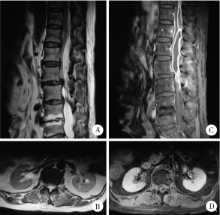
目的 总结原发性椎管内脓肿的特点以及治疗方法,以期提高原发性椎管内脓肿的诊断与治疗水平.方法: 回顾性分析北京大学第三医院近20年来收治的13例原发性椎管内脓肿的病例,分别对其病史,病因,病原学,手术方式以及预后进行分析.结果: 所有病例病程从7 d到6个月不等,所有病例均以疼痛起病,有10例自觉肢体无力,有5例患者有发热史,8例血常规白细胞升高.13例中颈椎管内脓肿1例,颈胸段1例,胸段1例,胸腰段4例,腰骶段6例.病原学中,细菌培养阳性4例,病理学诊断结核3例,1例因发病前感染布氏杆菌(Brucella)按该细菌感染治疗,其余病例病原学结果均阴性.所有病例均行手术治疗,并获得组织病理学诊断.手术目的主要为清除病灶并减压,引流.术后根据病原学结果及临床经验予以抗感染及激素治疗.有2例术后形成切口脓肿再次行手术治疗,手术为清创及转移肌瓣修复.随访0.5~3.0年,平均1.8年.随访期1例术后复发并沿椎管扩散,再次手术治疗.所有病例最后炎症均完全恢复,神经系统体征较术前均有不同程度好转.结论: 原发性椎管内脓肿发病相对较急,主要以疼痛起病,腰骶部最好发,细菌培养多以阴性为主.建议早期手术,使用广谱,足量抗生素.术后如出现切口脓肿形成,宜在清创基础上转移肌瓣修复.术后为缓解脊髓和神经根水肿,可在抗生素保障下使用激素.

关键词: 椎管内脓肿, 手术, 抗生素, 激素, 肌瓣修复

Abstract:

Objective: To summarize the feature and treatment of the primary intraspinal abscess in order to improve the prognosis.Methods: In the study, 13 cases of primary intraspinal abscess of the recent 20 years were retrospectively analyzed. The history, etiology, pathogen, surgical methods and prognosis were summarized.Results: The course of the illness ranged from 7 days to 6 months. All the cases began with pain. Of the 13 patients, 10 had limb weakness. Five had a fever and 8 had increased white blood cells. As for distribution, 1 was in cervical vertebra, 1 in cervicothoracic junction, 1 in thoracic vertebra, 4 in thoracolumbar junction, and 6 in lumbosacral segment. The results of bacterial culture were positive in 4 cases, 3 cases were diagnosed as tuberculosis by pathological examination, and 1 case was recognized as infection of Brucella melitensis bacteria because of prior brucellosis. The pathogen of the remaining cases were unclear. All the cases received surgical treatment and pathology examination. The surgical aim was mainly removal of the lesion, decompression and drainage. Postoperatively anti-infection and glucocorticoid therapy were performed according to the pathogeny results and clinical experience. Incision abscesses were seen in 2 cases and reoperations including debridement and repair with transferred muscle flap were performed. Postoperative follow-up ranged from 6 months to 3 years (mean 1.8 years). One case suffered postoperative recurrence and the abscess spread along the vertebral canal. Reoperation was performed. Infections of all the cases were recovered completely and the nervous system signs were all improved in different degrees.Conclusion: The onset of primary intraspinal abscess is relatively urgent, mainly with pain. The lumbar and sacral vertebra is the predilection site. The bacterial culture is mostly negative. Early operation and use of sufficient amount of broad-spectrum antibiotic are recommended. If the incision abscess forms after the operation, it is advisable to transfer the muscle flap to repair the coloboma on the basis of debridement. In order to relieve edema of spinal cord and nerve root, the glucocorticoid can be used in the escort of antibiotics.

Key words: Intraspinal abscess, Surgery, Antibiotics, Glucocorticoid, Muscle flap

中图分类号: 

  • R632.5

表1

患者一般资料"

Case Gender Age Predisposing factor Course/d Onset symptom Temperature Neurological signs
1 Female 13 Diabetes mellitus 30 Low back pain and lower extremity pain Normal Weakness of lower limb and loss of lower limb tendon reflex
2 Female 15 None 180 Right upper limb pain Normal Weakness and hypoesthesia in the right upper limb
3 Male 18 Malignancy 7 Low back pain and lower extremity pain Fever Local tenderness, hypoesthesia between T10 to L1 and hyperesthesia between L2-L5
4 Female 24 Long term use of glucocorticoid 7 Low back pain Fever Weakness of lower limb, hypoesthesia below T12, hyporeflexiaof lower limb tendon reflex and relaxation of anal sphincter
5 Male 30 History of brucellosis 20 Neck pain Normal Weakness of right limb, positive right Babinski's sign and hypoesthesia at C2
6 Female 34 None 180 Low back pain Fever Local tenderness, hypoesthesia of right lower limb, weakness of lower limb, hyporeflexia of lower limb tendon reflex and meningeal irritation sign
7 Female 46 Malignancy, Diabetes mellitus 18 Low back pain Normal Weakness of lower limb and hyporeflexia of lower limb tendon reflex
8 Female 51 Local dermal sinus 15 Low back pain Normal Weakness of lower limb, hyperreflexia of lower limb tendon reflex, hypermyotonia and hyperesthesia of lower limb
9 Male 54 Local dermal sinus 10 Low back pain Normal Weakness of lower limb, hyperreflexia of lower limb tendon reflex, hypermyotonia and hyperesthesia of lower limb
10 Male 56 None 14 Low back pain Fever Local tenderness, weakness of lower limb and meningeal irritation sign
11 Female 58 None 20 Chest back pain Normal Weakness of lower limb, hyperreflexia of lower limb tendon reflex, pathological signs and hypoesthesia below T6
12 Female 64 None 30 Lumbosacral pain Fever Local tenderness, weakness of lower limb and hyporeflexia of lower limb tendon reflex
13 Male 68 Adjacent skin infection 7 Low back pain Normal Paralysis of lower limbs and relaxation of anal sphincter

表2

患者术前辅助检查"

Case Inflammatory markers MRI
WBC ESR CRP
1 Normal High NA Subdural lesion between L4-S1 with significant enhancement
2 Normal High NA Epidural lesion between C7-T3 with area of cystoid variation and necrosis and significant enhancement of solid area
3 High NA NA Epidural lesion in the thoraco-lumbo-sacral spinal canal with posterior vertical spine muscle strip enhancement, posterior lamellar muscle enhancement
4 High NA NA Epidural cystic lesion between T12-L2 with enhancement of cyst wall
5 Normal Normal High Epidural lesion ventral to spinal cord between C2-C4 with significant homogeneous enhancement and edema of corresponding spinal cord
6 Normal NA NA Multiple subdural lesions between T12-L3 with significant homogeneous enhancement
7 Normal High NA Intraspinal cystic lesion between T8-L2 with ring enhancement of cyst wall
8 High NA NA Intramedullary abnormal signal between L2-L3 with ring enhancement, dermal sinus at L4-L5 connected to the spinal canal with tethered cord
9 High NA NA Intramedullary abnormal signal between L2-L3 with ring enhancement, dermal sinus at L2-L3 connected to the spinal canal
10 High NA NA Intraspinal lesion between L3-L5 with ring enhancement
11 High NA NA Extramedullary epidural lesion between T4-T5 with ring enhancement
12 High High High Multiple intraspinal and paravertebral cystic lesions between S1-S3 with ring enhancement
13 High NA NA Epidural cystic lesions between T9-L1 with ring enhancement

图1

胸腰段硬膜外脓肿患者的术前磁共振检查"

表3

治疗情况及术中所见"

Case Treatment Location Abscess character
Surgery Antibacterials Glucocorticoid Mannitol
1 Yes Yes(anti-TB) No No L4-S1, epidural, ventral Gelatinous and caseous
2 Yes Yes(anti-TB) No No C7-T3, epidural Thinning pus and gray granuloma
3 Yes Yes No No L4-S1, epidural Yellowish-white thinning pus
4 Yes Yes Yes No T12-L2, epidural Beige pus
5 Yes Yes Yes Yes C3-C7, epidural, ventral Purple-red granuloma
6 Yes Yes No No T12-L3,intra-extramedullary Yellow sticky pus and granuloma
7 Yes Yes Yes No T8-L2, epidural Much yellow pus with granuloma
8 Yes Yes Yes Yes L2-L3,intra-extramedullary Yellowish-white pus with much pus mosses
9 Yes Yes Yes Yes L2-L3,subdural Yellowish-white pus
10 Yes Yes No No L3-L5, epi-subdural Yellowish-white pus with pus mosses and granuloma
11 Yes Yes Yes Yes T4-T5, intramedullary Yellowish-white pus
12 Yes Yes No No S1-S3, epi-subdural and paravertebral Yellowish-white pus with pale yellow granuloma
13 Yes Yes No No T9-L1, epidural Beige pus with little granuloma and much pus mosses

表4

患者术后情况"

NA, not applicable.

Case Pyoculture Blood culture Pathology Possible way Outcome
1 Negative Negative Tuberculosis Hematogenous Complete recovery
2 Negative NA Tuberculosis Hematogenous Complete recovery
3 Negative NA NA Hematogenous Complete recovery
4 Staphylococcus aureus NA Pus Hematogenous No abscess or pain, light hypoesthesia, muscle strength grade IV, occasional urine incontinence
5 NA NA NA Hematogenous No abscess or pain, euesthesia, muscle strength grade Ⅳ
6 Negative NA Pus Hematogenous Recurrence 1 year later, complete recovery finally
7 Negative NA Tuberculosis Hematogenous No abscess or pain, euesthesia, muscle strength grade Ⅳ
8 Enterococcus faecalis NA Dermal sinus, necrosis
and granulation tissue
Direct spread Complete recovery
9 Escherichia coli NA Dermal sinus Direct spread Complete recovery
10 Negative NA Pus Hematogenous No abscess or pain, euesthesia, muscle strength grade Ⅳ
11 Negative NA Pus Hematogenous No abscess or pain, hypoesthesia, no provement in muscle strength
12 Negative Negative Pus Hematogenous Wound abscess which restored after debridement and repair with muscle flap, complete recovery finally
13 Staphylococcus aureus NA Pus Hematogenous Wound abscess which restored after debridement and repair with muscle flap. No abscess or pain, hypoesthesia, muscle strength grade Ⅳ, occasional urine incontinence finally

图2

胸腰段硬膜外脓肿患者的术后核磁"

[1] Honig A, Or O, Barzilay Y , et al. Spinal epidural abscess with a rapid course in young healthy infantry recruits with multiple skin lacerations[J]. J Clin Neurosci, 2016,31:127-132.
[2] Reihsaus E, Waldbaur H, Seeling W . Spinal epidural abscess: a meta-analysis of 915 patients[J]. Neurosurg Rev, 2000,23(4):175-205.
[3] Ersahin Y . Spinal epidural abscess: a meta-analysis of 915 patients[J]. Neurosurg Rev, 2001,24(2/3):156.
[4] Karikari IO, Powers CJ, Reynolds RM , et al. Management of a spontaneous spinal epidural abscess: a single-center 10-year experience[J]. Neurosurgery, 2009,65(5):919-924.
[5] Bartels RH, de Jong TR, Grotenhuis JA . Spinal subdural abscess[J]. J Neurosurg, 1992,76(2):307-311.
[6] Kirzner H, Oh YK, Lee SH . Intraspinal air: a CT finding of epidural abscess[J]. Am J Roentgenol, 1988,151(6):1217-1218.
[7] Farber SH, Murphy KR, Suryadevara CM , et al. Comparing outcomes of early, late, and non-surgical management of intraspinal abscess[J]. J Clin Neurosci, 2017,36:64-71.
[8] Kulkarni AG, Chu G, Fehlings MG . Pyogenic intradural abscess: a case report[J]. Spine, 2007,32(12):E354-E357.
[9] Thomé C, Krauss JK, Zevgaridis D , et al. Pyogenic abscess of the filum terminale. Case report[J]. J Neurosurg, 2001,95(Suppl 1):100-104.
[10] Darouiche RO . Spinal epidural absces[J]. N Engl J Med, 2006,355(19):2012-2020.
[11] Hanci M, Sarioglu AC, Uzan M , et al. Intramedullary tuberculous abscess: a case report[J]. Spine, 1996,21(6):766-769.
[12] Bingöl A, Yücemen N, Meço O . Medically treated intraspinal "Brucella" granuloma[J]. Surg Neurol, 1999,52(6):570-576.
[13] Kamat AS, Thango NS, Husein MB . Proteus mirabilis abscess involving the entire neural axis[J]. J Clin Neurosci, 2016,30:127-129.
[14] Al Barbarawi M, Khriesat W, Qudsieh S , et al. Management of intramedullary spinal cord abscess: experience with four cases, pathophysiology and outcomes[J]. Eur Spine J, 2009,18(5):710-717.
[15] Hindy J, Shelef I, Slovik Y , et al. Late prevertebral and spinal abscess following chemoradiation for laryngeal squamous cell carcinoma[J]. Case Rep Otolaryngol, 2014,2014:425724.
[16] Miyazaki M, Yoshiiwa T, Kodera R , et al. Clinical features of cervical pyogenic spondylitis and intraspinal abscess[J]. J Spinal Disord Tech, 2011,24(7):E57-E61.
[17] Ur-rahman N, El-bakry A, Jamjoom AB , et al. Atypical forms of spinal tuberculosis: case report and review of the literature[J]. Surg Neurol, 1999,51(6):602-607.
[18] Khalil JG, Nassr A, Diehn FE , et al. Thoracolumbosacral spinal subdural abscess: magnetic resonance imaging appearance and limited surgical management[J]. Spine, 2013,38(13):E844-E847.
[19] Narlawar RS, Shah JR, Pimple MK , et al. Isolated tuberculosis of posterior elements of spine: magnetic resonance imaging findings in 33 patients[J]. Spine, 2002,27(3):275-281.
[20] Tanriverdi T, Kizilkiliç O, Hanci M , et al. Atypical intradural spinal tuberculosis: report of three cases[J]. Spinal Cord, 2003,41(7):403-409.
[21] Hung PC, Wang HS, Wu CT , et al. Spinal intramedullary abscess with an epidermoid secondary to a dermal sinus[J]. Pediatr Neurol, 2007,37(2):144-147.
[22] ter Avest E, Uyttenboogaart M, Dorgelo J , et al. A patient with neck pain and fever. Combined prevertebral and intraspinal abscess in a patient with a de novo HIV infection[J]. Neth J Med, 2009,67(10):356-357.
[23] Vajramani GV, Nagmoti MB, Patil CS . Neurobrucellosis presenting as an intramedullary spinal cord abscess[J]. Ann Clin Microbiol Antimicrob, 2005,16(4):14.
[24] Dev R, Husain M, Gupta A , et al. MR of multiple intraspinal abscesses associated with congenital dermal sinus[J]. Am J Neuroradiol, 1997,18(4):742-743.
[25] Nagar VR, Springer JE, Salles S . Increased incidence of spinal abscess and substance abuse after implementation of state mandated prescription drug legislation[J]. Pain Med, 2015,16(10):2031-2035.
[26] Singh I, Rohilla S, Kumar P , et al. Spinal dorsal dermal sinus tract: An experience of 21 cases[J]. Surg Neurol Int, 2015,6(Suppl 17):S429-434.
[1] 步召德, 冯梦宇, 季科. 早期胃癌行前哨淋巴结导航手术的实践与思考[J]. 北京大学学报(医学版), 2026, 58(2): 239-243.
[2] 高加勒, 张忠涛. 局部进展期直肠癌精准治疗现状与展望[J]. 北京大学学报(医学版), 2026, 58(2): 247-250.
[3] 付浩, 申潞艳, 黄冰洋, 马少华. 免疫治疗背景下食管鳞状细胞癌围手术期治疗的临床思考[J]. 北京大学学报(医学版), 2026, 58(2): 266-271.
[4] 吴曼, 罗樱樱, 纪立农. 女性睾酮检测中假阳性问题及质谱法的确诊价值[J]. 北京大学学报(医学版), 2026, 58(2): 332-336.
[5] 李斌, 梁寒. 机器人胃癌根治术:研究进展与实践挑战[J]. 北京大学学报(医学版), 2026, 58(2): 416-422.
[6] FarinEbrahimi, 冯志强, FarazEbrahimi, 韩玮华, 于子杨, 贾宽宽, 安金刚. 上颌药物相关性颌骨坏死的不同分期手术治疗效果[J]. 北京大学学报(医学版), 2026, 58(1): 107-114.
[7] 潘莲菲, 李文静, 王瑞洋, 焦剑, 曹战强, 高丽, 释栋. 口服抗生素辅助牙周机械治疗对重度牙周炎的短期疗效及影响因素[J]. 北京大学学报(医学版), 2026, 58(1): 30-36.
[8] 唐仁韬, 杨流畅, 聂杰, 王晓燕. 无窦型与有窦型根管治疗后慢性根尖周炎根管外菌群的组成及差异[J]. 北京大学学报(医学版), 2026, 58(1): 43-49.
[9] 王晓林, 郭邵逸, 陈大召, 温锡杰, 华勇, 张亮, 张秦. 全髋关节置换术治疗系统性红斑狼疮继发股骨头缺血性坏死的随访研究[J]. 北京大学学报(医学版), 2025, 57(6): 1081-1088.
[10] 李浙民, 季加孚, 李国新, 李子禹, 步召德, 高翔宇, 董迪, 唐磊, 邢晓芳, 贾淑芹, 郭婷, 张连海, 陕飞, 季鑫, 王安强. 胃癌精准诊疗技术的创建与推广[J]. 北京大学学报(医学版), 2025, 57(5): 864-867.
[11] 李博闻, 张强, 孙益鑫. 儿童及青年漏斗胸患者Nuss术后发生脊柱侧弯的风险预测模型建立及验证[J]. 北京大学学报(医学版), 2025, 57(5): 941-946.
[12] 李宗瀚, 黄洋阅, 李宁, 李明磊, 宋宏程, 张潍平, 刘超. 国产单孔蛇形臂机器人手术系统在儿童肾盂成形术中的应用[J]. 北京大学学报(医学版), 2025, 57(4): 662-665.
[13] 张启鸣, 陈泽波, 田雨, 潘大猛, 刘磊, 张洪宪, 赵磊, 张树栋, 马潞林, 侯小飞. 机器人辅助腹腔镜移植肾切除术经验总结[J]. 北京大学学报(医学版), 2025, 57(4): 666-669.
[14] 左超, 王国立, 杨昆霖, 车新艳, 孟一森, 张凯. 前列腺体积不同的患者经尿道光纤铥激光前列腺剜除术的有效性及安全性比较[J]. 北京大学学报(医学版), 2025, 57(4): 711-716.
[15] 张铃福, 王港, 侯纯升, 崔龙, 王立新, 凌晓锋, 徐智. 腹腔镜下改良经胆囊管胆管引流术在胆石症治疗及胆道疾病诊断中的应用[J]. 北京大学学报(医学版), 2025, 57(4): 748-752.
Viewed
Full text


Abstract

Cited

  Shared   
  Discussed   
No Suggested Reading articles found!