北京大学学报(医学版) ›› 2021, Vol. 53 ›› Issue (1): 183-187. doi: 10.19723/j.issn.1671-167X.2021.01.027
CT能谱曲线在脊柱转移瘤和感染性病变中的鉴别诊断价值
袁源,郎宁,袁慧书( )
)
- 北京大学第三医院放射科,北京 100191
CT spectral curve in differentiating spinal tumor metastasis and infections
YUAN Yuan,LANG Ning,YUAN Hui-shu()
- Department of Radiology, Peking University Third Hospital, Beijing 100191, China
摘要:
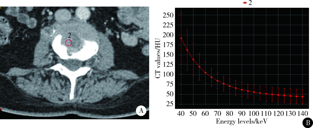
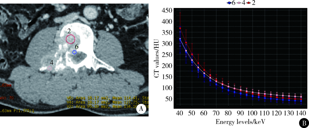
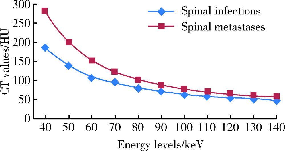
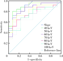
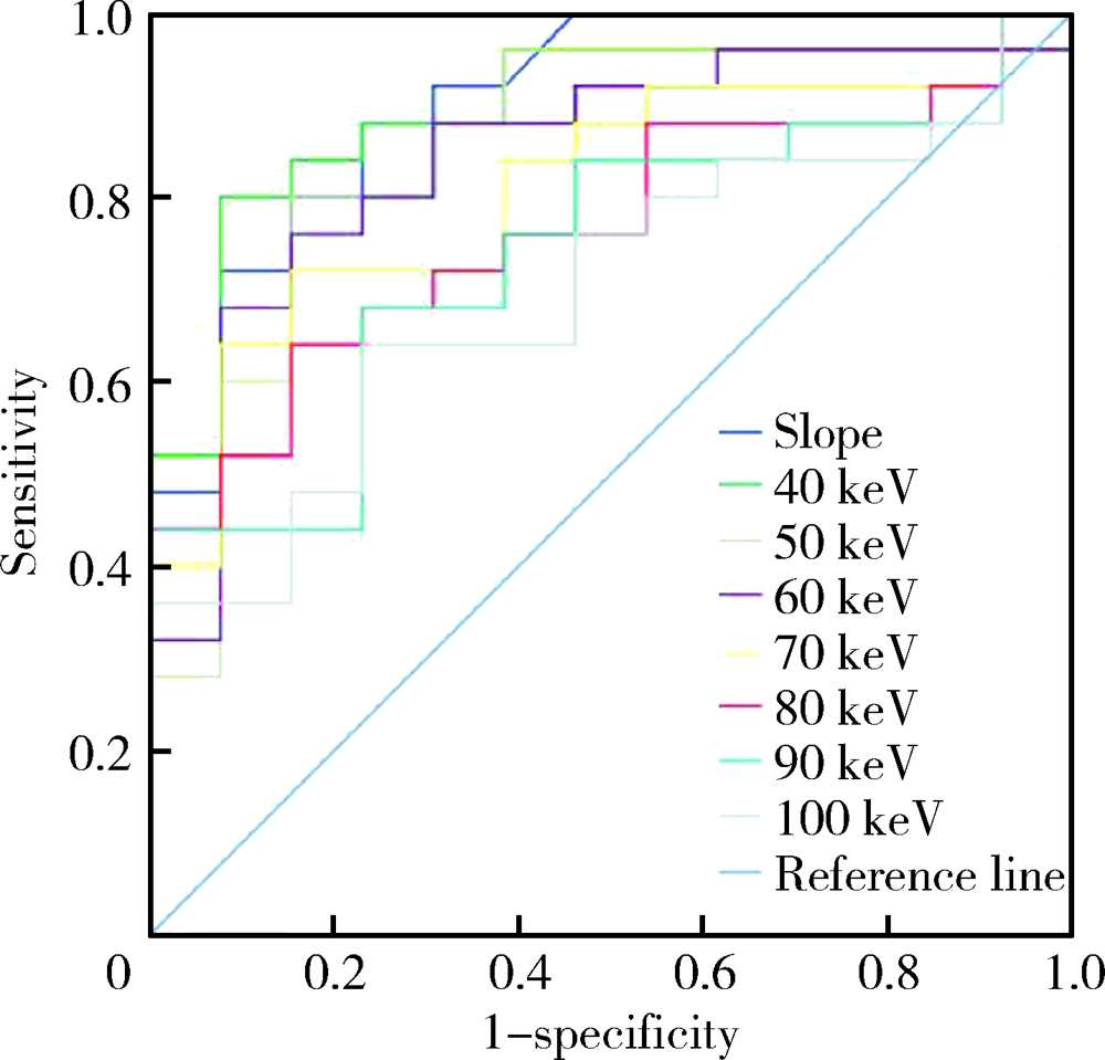
目的: 探讨CT能谱曲线在脊柱转移瘤和感染性病变中鉴别诊断的价值。方法: 收集经手术、穿刺组织病理检查或临床治疗证实的29例脊柱转移瘤和18例脊柱感染性病变的能谱CT增强静脉期扫描图像,经后处理获得单能量图像,生成病变感兴趣区(region of interest,ROI)CT能谱曲线,计算40~140 keV不同能级水平(每10 keV)下的CT值和能谱曲线斜率,同时在传统混合能量图像的同一ROI测量CT值,对所得CT值、能谱曲线斜率及其诊断效能进行统计学分析。结果: 脊柱转移瘤和感染性病变的中位年龄分别为58岁和64岁,两组差异无统计学意义(U=171,P=0.4)。40~100 keV各能级下脊柱转移瘤的CT值分别为281.79 (143.67, 446.19) HU、199.68 (100.04, 321.49) HU、151.54 (81.47, 243.49) HU、(122.64±27.72) HU、(99.90±23.88) HU、(85.82±21.61) HU、(75.94±20.27) HU,脊柱感染性疾病的CT值分别为185.29 (164.19, 277.03) HU、138.44 (124.98, 238.56) HU、105.46 (92.94, 169.53) HU、(93.77±15.55) HU、(79.15±12.84) HU、(68.99±11.75) HU、(62.22±11.71) HU。脊柱转移瘤在同一能级的CT值显著高于脊柱感染性病变(P均<0.05),110~140 keV能量水平下的CT值差异无统计学意义(P均>0.05)。混合能量图像测量的CT值差异无统计学意义(P>0.05)。脊柱转移瘤的能谱曲线斜率为2.43±0.58,高于脊柱感染性疾病的能谱曲线斜率1.50±0.40(P<0.001)。当静脉期能谱曲线斜率阈值取1.72、40 keV CT值取248.80 HU时,曲线下面积分别为0.905、0.892,灵敏度分别为88.0%、80.0%,特异度分别为76.9%、92.3%。结论: CT能谱曲线能够对鉴别脊柱转移瘤及感染性病变提供有价值的半定量信息,可以作为传统CT影像学的补充。
中图分类号:
- R738.1


| [1] |
Lee SH, Lee JM, Kim KW, et al. Dual-energy computed tomography to assess tumor response to hepatic radiofrequency ablation: potential diagnostic value of virtual noncontrast images and iodine maps[J]. Invest Radiol, 2011,46(2):77-84.
doi: 10.1097/RLI.0b013e3181f23fcd pmid: 20856125 |
| [2] |
Lang N, Yuan H, Yu HJ, et al. Diagnosis of spinal lesions using heuristic and pharmacokinetic parameters measured by dynamic contrast-enhanced MRI[J]. Acad Radiol, 2017,24(7):867-875.
doi: 10.1016/j.acra.2016.12.014 pmid: 28162875 |
| [3] |
Babic M, Simpfendorfer CS. Infections of the Spine[J]. Infect Dis Clin North Am, 2017,31(2):279-297.
doi: 10.1016/j.idc.2017.01.003 pmid: 28366222 |
| [4] |
Dong Y, Zheng S, Machida H, et al. Differential diagnosis of osteoblastic metastases from bone islands in patients with lung cancer by single-source dual-energy CT: advantages of spectral CT imaging[J]. Eur J Radiol, 2015,84(5):901-907.
doi: 10.1016/j.ejrad.2015.01.007 pmid: 25661696 |
| [5] |
Ko JP, Brandman S, Stember J, et al. Dual-energy computed tomography: concepts, performance, and thoracic applications[J]. J Thorac Imaging, 2012,27(1):7-22.
doi: 10.1097/RTI.0b013e31823fe0e9 pmid: 22189245 |
| [6] |
Flais J, Coiffier G, Brillet E, et al. Atypical presentation of spine bone metastasis in prostate cancer mimicking Pott’s disease[J]. Clin Cases Miner Bone Metab, 2017,14(2):239-240.
doi: 10.11138/ccmbm/2017.14.1.239 pmid: 29263741 |
| [7] | 袁源, 张艳, 郎宁, 等. CT能谱曲线鉴别诊断脊柱肿瘤及肿瘤样病变[J]. 中国医学影像技术, 2015,31(4):600-603. |
| [8] |
Avrin DE, Macovski A, Zatz LE. Clinical application of Compton and photo-electric reconstruction in computed tomography: preliminary results[J]. Invest Radiol, 1978,13(3):217-222.
doi: 10.1097/00004424-197805000-00007 pmid: 711396 |
| [9] | Dilmanian FA. Computed tomography with monochromatic X rays[J]. Am J Physiol Imaging, 1992,7(3/4):175-193. |
| [10] |
Riederer SJ, Mistretta CA. Selective iodine imaging using K-edge energies in computerized X-ray tomography[J]. Med Phys, 1977,4(6):474-481.
doi: 10.1118/1.594357 pmid: 927384 |
| [11] |
Silva AC, Morse BG, Hara AK, et al. Dual-energy (spectral) CT: applications in abdominal imaging[J]. Radiographics, 2011,31(4):1031-1050.
doi: 10.1148/rg.314105159 pmid: 21768237 |
| [12] | 雷立昌, 陈建宇. 能谱CT的临床应用与研究进展[J]. 中国医学影像技术, 2013,29(1):146-149. |
| [13] | 林晓珠, 沈云, 陈克敏. CT能谱成像的基本原理与临床应用研究进展[J]. 中华放射学杂志, 2011,45(8):798-800. |
| [14] | 张靖, 周新社. 脊柱肿瘤的诊断和外科分期研究进展[J]. 中华全科医学, 2011,9(2):277-279. |
| [15] |
Go SW, Lee HY, Lim CH, et al. Atypical disseminated skeletal tuberculosis mimicking metastasis on PET-CT and MRI[J]. Intern Med, 2012,51(20):2961-2965.
doi: 10.2169/internalmedicine.51.8347 pmid: 23064577 |
| [16] |
Mittal S, Khalid M, Sabir AB, et al. Comparison of magnetic resonance imaging findings between pathologically proven cases of atypical tubercular spine and tumour metastasis: A retrospective study in 40 patients[J]. Asian Spine J, 2016,10(4):734-743.
doi: 10.4184/asj.2016.10.4.734 pmid: 27559455 |
| [17] |
Sezgin B, Atilganoglu U, Yigit O, et al. Concomitant cutaneous metastatic tuberculous abscesses and multifocal skeletal tuberculosis[J]. Indian J Dermatol, 2008,53(3):149-153.
doi: 10.4103/0019-5154.43208 pmid: 19882018 |
| [18] |
Chang DS, Rafii M, McGuinness G, et al. Primary multifocal tuberculous osteomyelitis with involvement of the ribs[J]. Skeletal Radiol, 1998,27(11):641-645.
doi: 10.1007/s002560050451 pmid: 9867183 |
| [19] |
Lang N, Su MY, Yu HJ, et al. Differentiation of tuberculosis and metastatic cancer in the spine using dynamic contrast-enhanced MRI[J]. Eur Spine J, 2015,24(8):1729-1737.
doi: 10.1007/s00586-015-3851-z pmid: 25749725 |
| [20] |
Zheng S, Dong Y, Miao Y, et al. Differentiation of osteolytic metastases and Schmorl’s nodes in cancer patients using dual-energy CT: Advantage of spectral CT imaging[J]. Eur J Radiol, 2014,83(7):1216-1221.
doi: 10.1016/j.ejrad.2014.02.003 pmid: 24820064 |
| [21] | Gupta S, Wagner-Bartak N, Jensen CT, et al. Dual-energy CT of pancreatic adenocarcinoma: Reproducibility of primary tumor measurements and assessment of tumor conspicuity and margin sharpness[J]. Abdom Radiol (NY), 2016,41(7):1317-1324. |
| [22] |
Ramon A, Bohm-Sigrand A, Pottecher P, et al. Role of dual-energy CT in the diagnosis and follow-up of gout: systematic analysis of the literature[J]. Clin Rheumatol, 2018,37(3):587-595.
doi: 10.1007/s10067-017-3976-z pmid: 29350330 |
| [1] | 马豆豆, 马晓彩, 常天静, 王丽芳, 丁艳, 石连杰. 临床表现似系统性红斑狼疮的大B细胞淋巴瘤骨髓受累1例[J]. 北京大学学报(医学版), 2026, 58(3): 666-669. |
| [2] | 赵业, 刁小莉, 熊焰. 细胞转移技术在微量细胞液病理诊断中的应用[J]. 北京大学学报(医学版), 2026, 58(1): 208-213. |
| [3] | 王月, 梁宇红. 繁茂型牙骨质-骨结构不良1例[J]. 北京大学学报(医学版), 2026, 58(1): 220-224. |
| [4] | 池彦廷, 蒋鸿杰, 陈艳, 徐志秀, 李斌斌. 直接免疫荧光在口腔黏膜寻常型天疱疮诊断中的价值: 基于多指标联合分析的回顾性研究[J]. 北京大学学报(医学版), 2026, 58(1): 68-73. |
| [5] | 顾静妍, 李欣艺, 赵金霞, 穆荣. 误诊为类风湿关节炎、痛风的糖尿病致Charcot关节病1例[J]. 北京大学学报(医学版), 2025, 57(6): 1193-1197. |
| [6] | 肖晓笛, 夏有辰, 柳剑英, 付鹏. 左侧胸锁乳突肌间血管内乳头状内皮增生1例[J]. 北京大学学报(医学版), 2025, 57(5): 1002-1004. |
| [7] | 孙翔宇, 袁超, 周芯竹, 刁婧, 郑树国. 唾液微生态在口腔及全身疾病早期防治中的应用[J]. 北京大学学报(医学版), 2025, 57(5): 859-863. |
| [8] | 叶嘉慧, 王时敏, 王子轩, 刘云松, 孙玉春, 叶红强, 周永胜. 基于CBCT图像构建牙颌面虚拟患者的两种配准方法比较[J]. 北京大学学报(医学版), 2025, 57(2): 354-359. |
| [9] | 陈钊, 邱永康, 康磊. 经典型Sweet综合征 18F-FDG PET/CT多脏器异常显像1例[J]. 北京大学学报(医学版), 2025, 57(2): 403-407. |
| [10] | 方媛媛, 徐帆, 雷杰, 张昊, 张文宇, 孙宇, 吴宏新, 傅开元, 毛伟玉. 基于颞下颌关节紊乱病诊断标准的临床自动诊断系统的建立及验证[J]. 北京大学学报(医学版), 2025, 57(1): 192-201. |
| [11] | 王昕莹, 程雪原, 张勇, 李菲, 段晋瑜, 乔静. 浓缩生长因子与自固化磷酸钙人工骨联合治疗牙周骨下袋缺损的疗效:临床和影像学评价[J]. 北京大学学报(医学版), 2025, 57(1): 42-50. |
| [12] | 车佳璐, 刘子臣, 李琨, 张晨, 车南颖. 全自动EasyNAT核酸快速检测系统检测石蜡包埋组织诊断结核病的临床价值[J]. 北京大学学报(医学版), 2024, 56(6): 1047-1051. |
| [13] | 陈心心, 唐哲, 乔艳春, 荣文笙. 北京市密云区4岁儿童患龋状况及其与龋活跃性检测的相关性[J]. 北京大学学报(医学版), 2024, 56(5): 833-838. |
| [14] | 钟华, 李原, 徐丽玲, 白明欣, 苏茵. 18F-FDG PET/CT在风湿免疫病中的应用[J]. 北京大学学报(医学版), 2024, 56(5): 853-859. |
| [15] | 李正芳,罗采南,武丽君,吴雪,孟新艳,陈晓梅,石亚妹,钟岩. 抗氨基甲酰化蛋白抗体在诊断类风湿关节炎中的应用价值[J]. 北京大学学报(医学版), 2024, 56(4): 729-734. |
|
||
