Journal of Peking University(Health Sciences) ›› 2018, Vol. 50 ›› Issue (6): 1083-1087. doi: 10.19723/j.issn.1671-167X.2018.06.025
• Article • Previous Articles Next Articles
Staining effect of bovine incisors in vitro by tea solutions
Meng-meng ZHU1,Ying-long LI2,Jie PAN1,△( )
)
- 1. Department of General, Peking University School and Hospital of Stomatology & National Clinical Research Center for Oral Diseases & National Engineering Laboratory for Digital and Material Technology of Stomatology & Beijing Key Laboratory of Digital Stomatology, Beijing 100081, China
2. Academy for Advanced Interdisciplinary Studies, Peking University, Beijing 100871, China
CLC Number:
- R781

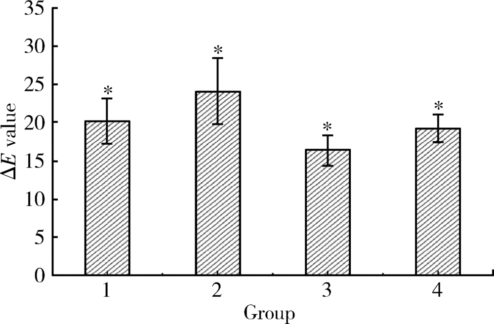

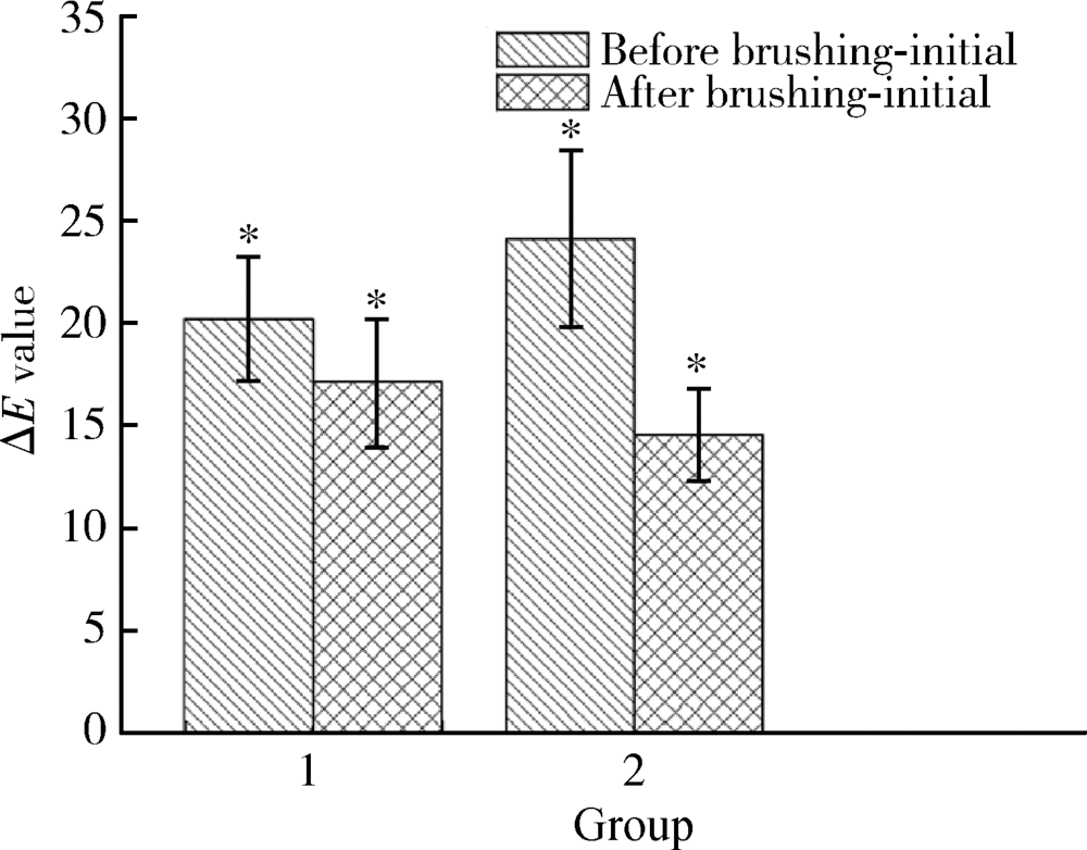
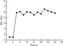

| [1] |
Rajesh AG, Ranganath LM, Kumar KS , et al. Surface morpho-logical changes in human enamel following bleaching: an in vitro scanning electron microscopic study[J]. J Contemp Dent Pract, 2012,13(3):405-415.
doi: 10.5005/jp-journals-10024-1159 pmid: 22918018 |
| [2] |
Xing W, Jiang T, Liang S , et al. Effect of in-office bleaching agents on the color changes of stained ceromers and direct com-posite resins[J]. Acta Odontol Scand, 2014,72(8):1032-1038.
doi: 10.3109/00016357.2014.946962 pmid: 25196263 |
| [3] |
Lee BS, Huang SH, Chiang YC , et al. Development of in vitro tooth staining model and usage of catalysts to elevate the effectiveness of tooth bleaching[J]. Dent Mater, 2008,24(1):57-66.
doi: 10.1016/j.dental.2007.01.012 pmid: 17442388 |
| [4] |
Lee RJ, Bayne A, Tiangco M , et al. Prevention of tea-induced extrinsic tooth stain[J]. Int J Dent Hyg, 2014,12(4):267-272.
doi: 10.1111/idh.12096 pmid: 25040739 |
| [5] |
Agnihotry A, Gill KS, Singhal D , et al. A Comparison of the bleaching effectiveness of chlorine dioxide and hydrogen peroxide on dental composite[J]. Braz Dent J, 2014,25(6):524-527.
doi: 10.1590/0103-6440201300098 pmid: 25590199 |
| [6] |
Homayoom A, Sadat HKS, Nafiseh Z , et al. In vitro effect of casein phosphopeptide amorphous calcium phosphate on enamel susceptibility to staining by tea during bleaching treatment[J]. J Dent, 2015,12(8):607-613.
pmid: 27123021 |
| [7] |
Çelik C, Yüzügüllü B, Erkut S , et al. Effect of bleaching on staining susceptibility of resin composite restorative materials[J]. J Esthet Restor Dent, 2010,21(6):407-414.
doi: 10.1111/j.1708-8240.2009.00299.x pmid: 20002929 |
| [8] |
Suliemana M, Addya M, Macdonalda E , et al. The bleaching depth of a 35% hydrogen peroxide based in-office product: a study in vitro[J]. J Dent, 2005,33(1):33-40.
doi: 10.1016/j.jdent.2004.07.008 |
| [9] |
Sulieman M, Addy M, Rees JS . Development and evaluation of a method in vitro to study the effectiveness of tooth bleaching[J]. J Dent, 2003,31(6):415-422.
doi: 10.1016/S0300-5712(03)00069-1 pmid: 12878024 |
| [10] | 路丽 . 着色牙外漂白后再矿化的效果及对细菌粘附影响的体外实验研究[D]. 山东: 山东大学, 2005. |
| [11] | 李艳 . 三种着色牙模型漂白前后颜色的比较研究[D]. 湖南: 中南大学, 2012. |
| [12] |
Odaira C, Itoh S, Ishibashi K . Clinical evaluation of a dental color analysis system: the crystaleyespectrophotometer[J]. J Prosthodont Res, 2011,55(4):199-205.
doi: 10.1016/j.jpor.2010.12.005 pmid: 21296639 |
| [13] |
Wang C, Li Y, Wang X , et al. The enamel microstructures of bovine Omandibular incisors[J]. Anat Rec, 2012,295(10):1698-1706.
doi: 10.1002/ar.22543 pmid: 22837182 |
| [14] |
Addy M, Moran J . Mechanism of stain formation on teeth, in particular associated with metal ions and antiseptics[J]. Adv Dent Res, 1995,9(4):450-456.
doi: 10.1177/08959374950090041601 |
| [15] |
Carpenter GH, Pramanik R, Proctor GB . An in vitro model of chlorhexidine-induced tooth staining[J]. J Periodental Res, 2005,40(3):225-230.
doi: 10.1111/j.1600-0765.2005.00791.x pmid: 15853968 |
| [16] |
Berger SB, Coelho AS, Oliveira VA , et al. Enamel susceptibility to red wine staining after 35% hydrogen peroxide bleaching[J]. J Appl Oral Sci, 2008,16(3):201-204.
doi: 10.1590/S1678-77572008000300007 |
| [17] |
Phan X, Akyalcin S, Wiltshire WA , et al. Effect of tooth bleaching on shear bond strength of a fluoride- releasing sealant[J]. Angle Orthod, 2012,82(3):546-551.
doi: 10.2319/052711-353.1 pmid: 21854189 |
| [18] |
Joiner A, Muller D, Elofsson UM , et al. Ellipsometry analysis of the in vitro adsorption of tea polyphenols onto salivary pellicles[J]. Eur J Oral Sci, 2004,112(6):510-515.
doi: 10.1111/j.1600-0722.2004.00166.x pmid: 15560834 |
| [19] |
Pontefract H, Courtney M, Smith S , et al. Development of methods to enhance extrinsic tooth discoloration for comparison of toothpastes I studies in vitro[J]. J Clin Periodontol, 2004,31(1):1-6.
doi: 10.1111/cpe.2004.31.issue-1 |
| [20] |
Li Q, Xu BT, Li R , et al. Quantitative evaluation of colour regression and mineral content change of bleached teeth[J]. J Dent, 2010,38(3):253-260.
doi: 10.1016/j.jdent.2009.11.005 pmid: 19945498 |
| [21] |
李青, 王贻宁, 徐东选 . 四种常见离体牙保存液对牙齿颜色的影响. 口腔医学研究, 2005,21(2):197-199.
doi: 10.3969/j.issn.1671-7651.2005.02.027 |
| [1] | Jing YANG, Xiaoyun XU, Danni ZHENG, Xiaotong LING, Liuyang QU, Denggao LIU. Clinical and imaging characteristics and etiology of 544 cases with chronic sialadenitis [J]. Journal of Peking University (Health Sciences), 2026, 58(3): 650-657. |
| [2] | Xinying WANG, Xueyuan CHENG, Mengjun ZHANG, Fei LI, Jinyu DUAN, Jing QIAO. Effect of concentrated growth factors in guided tissue regeneration for the treatment of mandibular molar furcation lesions [J]. Journal of Peking University (Health Sciences), 2026, 58(2): 372-379. |
| [3] | Xin CONG, Jiazeng SU, Liling WU, Chong DING, Wei LI, Guangyan YU. Research progress in diagnosis and treatment of non-tumorous salivary gland diseases [J]. Journal of Peking University (Health Sciences), 2026, 58(1): 1-9. |
| [4] | Yuting YANG, Liuyang QU, Danni ZHENG, Xiaotong LING, Xiaoyun XU, Denggao LIU. Demographic characteristic and clinical features in 1 812 patients with salivary gland stones [J]. Journal of Peking University (Health Sciences), 2026, 58(1): 153-159. |
| [5] | Hailing ZANG, Yuhong LIANG. Endodontic retreatment of a maxillary second molar with chronic apical periodontitis and separated instrument: A case report [J]. Journal of Peking University (Health Sciences), 2026, 58(1): 214-219. |
| [6] | Lianfei PAN, Wenjing LI, Ruiyang WANG, Jian JIAO, Zhanqiang CAO, Li GAO, Dong SHI. Short-term efficacy and influencing factors of systemic antibiotics as an adjunct to mechanical periodontal therapy for stages Ⅲ/Ⅳ periodontitis [J]. Journal of Peking University (Health Sciences), 2026, 58(1): 30-36. |
| [7] | Fei SUN, Cui WANG, Siqi LI, Yiping WEI, Riyue YU, Wenjie HU. Treatment of peri-implant mucositis using an erythritol air-polishing or ultrasonic device: A randomized controlled trial [J]. Journal of Peking University (Health Sciences), 2026, 58(1): 37-42. |
| [8] | Rentao TANG, Liuchang YANG, Jie NIE, Xiaoyan WANG. Microbial communities in extraradicular infections of post-treatment apical periodontitis without or with sinus tracts [J]. Journal of Peking University (Health Sciences), 2026, 58(1): 43-49. |
| [9] | Ziyu HE, Hui ZHANG, Zhibin CHEN, Haixia XING, Jie PAN. Isolation, identification, and metabolic characterization of a Veillonella parvula isolated from supragingival plaque in a patient with rampant caries [J]. Journal of Peking University (Health Sciences), 2026, 58(1): 50-59. |
| [10] | Yanting CHI, Hongjie JIANG, Yan CHEN, Zhixiu XU, Binbin LI. Value of direct immunofluorescence in the diagnosis of oral mucosal pemphigus vulgaris: A retrospective study based on multi-index combined analysis [J]. Journal of Peking University (Health Sciences), 2026, 58(1): 68-73. |
| [11] | Baojin MA, Jianhua LI, Yuanhua SANG, Yang YU, Jichuan QIU, Jinlong SHAO, Kai LI, Shiyue LIU, Mi DU, Lingling SHANG, Shaohua GE. Establishment and application of key technologies for periodontal tissue regeneration based on microenvironment and stem cell regulation [J]. Journal of Peking University (Health Sciences), 2025, 57(5): 841-846. |
| [12] | Pei CAO, Qingxian LUAN. Periodontitis and systemic diseases: Thinking and explorations [J]. Journal of Peking University (Health Sciences), 2025, 57(5): 852-858. |
| [13] | Xiangyu SUN, Chao YUAN, Xinzhu ZHOU, Jing DIAO, Shuguo ZHENG. Application of salivary micro-ecosystem in early prevention and control of oral and systemic diseases [J]. Journal of Peking University (Health Sciences), 2025, 57(5): 859-863. |
| [14] | Zhenying BAO, Yajie WANG. Application of combined detection of inflammatory indexes and cytokines in chronic periodontitis [J]. Journal of Peking University (Health Sciences), 2025, 57(4): 772-778. |
| [15] | 朱慧, 闵赛南, 苏家增, 陈艳, 彭歆, 于尧, 俞光岩. [J]. Journal of Peking University (Health Sciences), 2025, 57(3): 620-625. |
|
||