北京大学学报(医学版) ›› 2026, Vol. 58 ›› Issue (3): 650-657. doi: 10.19723/j.issn.1671-167X.2026.03.027

• 论著 • 上一篇    下一篇

544例慢性唾液腺炎的临床与影像学特点及病因分析

杨静, 许晓韵, 郑丹妮, 凌晓彤, 屈留洋, 柳登高*()   

  1. 北京大学口腔医学院·口腔医院医学影像科,国家口腔医学中心,国家口腔疾病临床医学研究中心,口腔生物材料和数字诊疗装备国家工程研究中心,口腔数字医学北京市重点实验室,国家卫生健康委员会口腔医学计算机应用工程技术研究中心,国家药品监督管理局口腔生物材料重点实验室,北京 100081
  • 收稿日期:2024-04-18 出版日期:2026-06-18 发布日期:2025-03-07
  • 通讯作者: 柳登高

Clinical and imaging characteristics and etiology of 544 cases with chronic sialadenitis

Jing YANG, Xiaoyun XU, Danni ZHENG, Xiaotong LING, Liuyang QU, Denggao LIU*()   

  1. Department of Oral and Maxillofacial Radiology, Peking University School and Hospital of Stomatology & National Center for Stomatology & National Clinical Research Center for Oral Diseases & National Engineering Research Center of Oral Biomaterials and Digital Medical Devices & Beijing Key Laboratory of Digital Stomatology & NHC Research Center of Engineering and Technology for Computerized Dentistry & NMPA Key Laboratory for Dental Materials, Beijing 100081, China
  • Received:2024-04-18 Online:2026-06-18 Published:2025-03-07
  • Contact: Denggao LIU

RICH HTML


摘要:

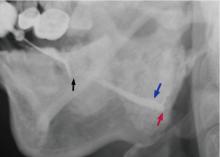
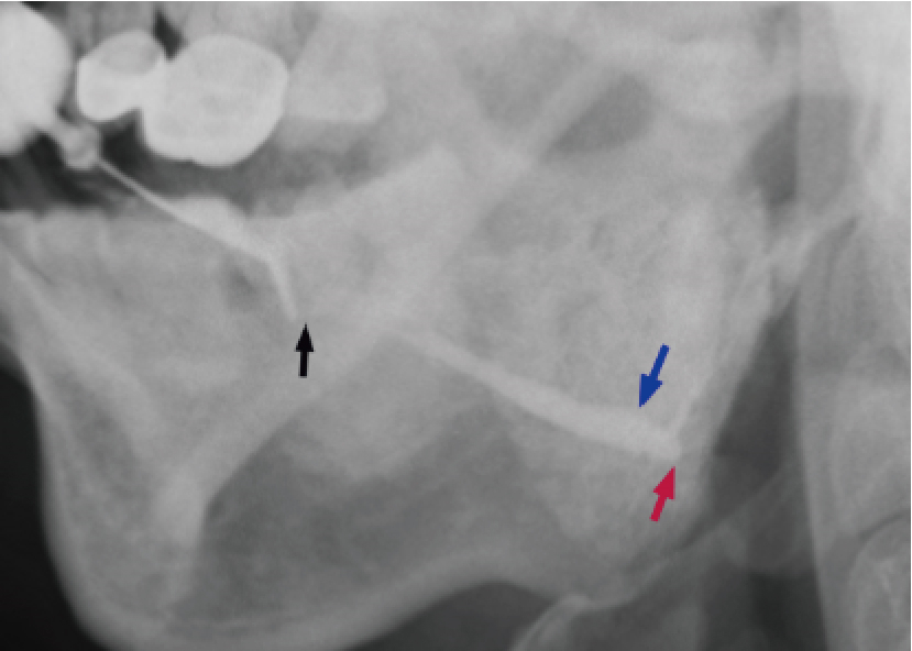
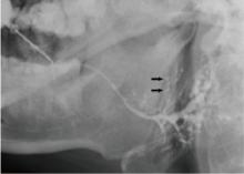
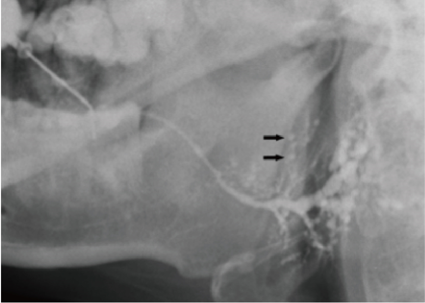
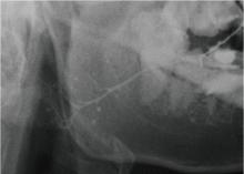
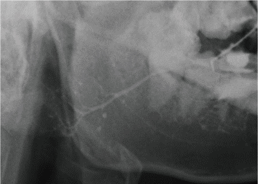
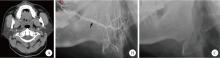
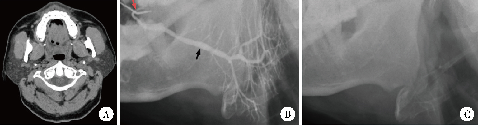
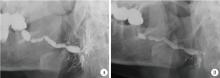
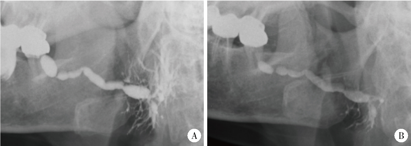
目的: 探索临床上常见的几类不同病因导致的慢性唾液腺炎的占比,并分析其临床和影像学特点。方法: 回顾性分析2021年1月至2023年8月就诊于北京大学口腔医院并行唾液腺内镜治疗的各类慢性唾液腺炎患者的临床及影像资料,除外干燥综合征伴感染、IgG4相关唾液腺炎、结石病伴感染等,根据现有研究结果和资料,初步将唾液腺炎分为131I相关唾液腺炎、过敏相关唾液腺炎、成人复发性腮腺炎、腮腺良性肥大伴管炎、原发性导管狭窄性唾液腺炎(指未发现目前已知的可能病因,并存在导管狭窄的慢性唾液腺炎),统计分析各类慢性唾液腺炎的占比和主要临床、影像学特点。结果: 共纳入544例患者,男性192例、女性352例,平均年龄(47.44±13.52)岁(14~83岁)。其中,原发性导管狭窄性唾液腺炎最多,占71.7%(390例);过敏相关唾液腺炎次之,占12.5%(68例);131I相关唾液腺炎占6.4%(35例),成人复发性腮腺炎占4.8%(26例),腮腺良性肥大伴管炎占4.6%(25例)。544例患者共1 120侧腺体受累,包括腮腺880侧(78.6%)及下颌下腺240侧(21.4%)。这五类唾液腺炎在性别、年龄、受累腺体类别、受累腺体数目及病程上差异均存在统计学意义(P < 0.05),131I相关唾液腺炎患者的女性占比最高(男∶女=1 ∶ 4.83),成人复发性腮腺炎患者的平均年龄最小[(32.50±8.60)岁],131I相关唾液腺炎和过敏相关唾液腺炎的受累腺体数目相对较多。从造影表现看,131I相关唾液腺炎中23.2%的受累腺体发生导管闭锁,过敏相关唾液腺炎中46.5%的受累腺体分支导管呈“雪花样”扩张表现,成人复发性腮腺炎中80.4%的受累腺体可见末梢导管“点球状扩张”,腮腺良性肥大伴管炎的受累腺体分支导管呈丛簇状表现者占71.4%,原发性导管狭窄性唾液腺炎的造影表现以主导管(主导管和分支导管)狭窄和(或)扩张为主。结论: 原发性导管狭窄性唾液腺炎、131I相关唾液腺炎、过敏相关唾液腺炎、成人复发性腮腺炎、腮腺良性肥大伴管炎是临床上常见的五类慢性唾液腺炎,其中原发性导管狭窄性唾液腺炎占比最大。明确各类慢性唾液腺炎的病因、临床及影像学特征,将有利于临床医师制定个性化治疗方案,改善治疗效果。

关键词: 唾液腺炎, 碘放射性同位素, 超敏反应, 腮腺, 下颌下腺

Abstract:

Objective: To investigate the etiology of chronic sialadenitis, and to analyze their clinical and imaging characteristics. Methods: This retrospective analysis reviewed the clinical and imaging data of patients with chronic sialadenitis who underwent sialendoscopy at the Peking University Hospital of Stomatology between January 2021 and August 2023. Inclusion criteria: (1) with a history of recurrent swelling of major salivary glands; (2) complete medical records with detailed information about potential causes; (3) sialography images were available; and (4) patients had undergone interventional endoscopy. Exclusion criteria: (1) salivary stones; (2) juvenile recurrent parotitis; (3) IgG4-related sialadenitis; (4) Sjögren syndrome; and (5) neoplastic diseases. Based on the latest research results and clinical data, chronic sialadenitis was classified into radioactive iodine-induced sialadenitis (RAIS), allergy-related sialodochitis (ARS), adult chronic recurrent parotitis (ACRP), sialadenosis with sialodochitis, and idiopathic sialadenitis. Idiopathic sialadenitis was defined as a type of chronic sialadenitis with duct stenosis of unknown etiology (allergic causes, autoimmune disorders, radioactive iodine exposure, history of "parotitis" in childhood, etc.). The proportions of five types of sialadenitis were calculated, and their relationships with age, gender, type of affected glands, number of affected glands, duration of symptoms, and imaging characteristics were statistically analyzed. Results: A total of 544 consecutive patients diagnosed with chronic sialadenitis were enrolled in this study. They consisted of 192 males and 352 females, and their ages ranged from 14 to 83 years [mean age: (47.44±13.52) years]. Idiopathic sialadenitis accounted for the largest proportion (71.7%, 390 cases), followed by ARS (12.5%, 68 cases), RAIS (6.4%, 35 cases), ACRP (4.8%, 26 cases), and sialadenosis with sialodochitis (4.6%, 25 cases). Among the 2 176 available glands of the 544 patients, 1 120 (51.5%) glands were affected, including 880 (78.6%) parotid glands and 240 (21.4%) submandibular glands. These five types of sialadenitis exhibited significant differences in gender, age, type and number of affected glands, and duration of symptoms (P < 0.05). Among them, RAIS patients showed the lowest male to female ratio (male ∶ female=1 ∶ 4.83), ARCP patients were the youngest [(32.50±8.60) years], and RAIS and ARS patients had relatively higher number of affected glands. Sialography showed ductal atresia in 23.2% of affected glands with ARIS, "snowflake" pattern in 46.5% of affected glands with ARS, "punctate and globular" ectasia of terminal ducts in 80.4% of affected glands with ARCP, and clustered branch ducts in 71.4% of affected glands with sialadenosis with sialodochitis. Moreover, stenosis and/or dilatation of the main and branch ducts represented the most typical sialography appearance of idiopathic sialadenitis. Conclusion: Idiopathic sialadenitis, RAIS, ARS, ACRP, and sialadenosis with sialodochitis are the five common types of chronic sialadenitis. Among these, idiopathic sialadenitis is the most common type of chronic sialadenitis. Clarification of the etiology, clinical manifestations and imaging characteristics of chronic sialadenitis is important for clinicians to develop personalized treatment plans and improve treatment outcomes.

Key words: Sialadenitis, Iodine radioisotopes, Hypersensitivities, Parotid gland, Submandibular gland

中图分类号: 

  • R781.72

图1

131I相关唾液腺炎的造影图像"

图2

过敏相关唾液腺炎的造影图像"

图3

成人复发性腮腺炎的造影图像"

图4

腮腺良性肥大伴管炎的造影及CT图像"

图5

原发性导管狭窄性唾液腺炎的造影图像"

表1

五类慢性唾液腺炎的临床特征"

Item Total (n=544) Idiopathic sialadenitis (n=390) ARS (n=68) RAIS (n=35) ACRP (n=26) Sialadenosis (n=25) P value
Gender, n(%) 0.025
  Male 192 (35.3) 137 (35.1) 23 (33.8) 6 (17.1) 12 (46.2) 14 (56.0)
  Female 352 (64.7) 253 (64.9) 45 (66.2) 29 (82.9) 14 (53.8) 11 (44.0)
Age/years, $\bar x \pm s$ 47.44±13.52 49.15±13.47 46.16±11.82 39.71±10.56 32.50±8.60 50.72±13.36 < 0.001
Type of affected glands, n(%) < 0.001
  PG only 417 (76.7) 319 (81.8) 28 (41.2) 19 (54.3) 26 (100.0) 25 (100.0)
  SMG only 73 (13.4) 53 (13.6) 20 (29.4) 0 (0.0) 0 (0.0) 0 (0.0)
  PG & SMG 54 (9.9) 18 (4.6) 20 (29.4) 16 (45.7) 0 (0.0) 0 (0.0)
Number of affected glands, n(%) < 0.001
  1 68 (12.5) 61 (15.6) 3 (4.4) 2 (5.7) 1 (3.8) 1 (4.0)
  2 424 (77.9) 313 (80.3) 45 (66.2) 17 (48.6) 25 (96.2) 24 (96.0)
  3 or 4 52 (9.6) 16 (4.1) 20 (29.4) 16 (45.7) 0 (0.0) 0 (0.0)
Duration of symptoms/years, n(%) < 0.001
  ≤1 282 (51.8) 215 (55.1) 21 (30.9) 30 (85.7) 0 (0.0) 16 (64.0)
  >1 & ≤10 205 (37.7) 148 (37.9) 42 (61.8) 5 (14.3) 2 (7.7) 8 (32.0)
  >10 57 (10.5) 27 (6.9) 5 (7.4) 0 (0.0) 24 (92.3) 1 (4.0)
1
Vashishta R , Gillespie MB . Salivary endoscopy for idiopathic chronic sialadenitis[J]. Laryngoscope, 2013, 123 (12): 3016- 3020.

doi: 10.1002/lary.24211
2
Erdoǧan O , Özcan C , İsmi O , et al. Effectiveness of sialendoscopy on the symptoms of chronic obstructive sialadenitis and patient satisfaction degree[J]. J Stomatol Oral Maxillofac Surg, 2022, 123 (3): 314- 319.

doi: 10.1016/j.jormas.2021.05.010
3
Pace CG , Hwang KG , Papadaki M , et al. Interventional sialoendoscopy for treatment of obstructive sialadenitis[J]. J Oral Maxillofac Surg, 2014, 72 (11): 2157- 2166.

doi: 10.1016/j.joms.2014.06.438
4
Sánchez Barrueco á , Santillán Coello JM , González Galán F , et al. Correction to: Epidemiologic, radiologic, and sialendoscopic aspects in chronic obstructive sialadenitis[J]. Eur Arch Otorhinolaryngol, 2023, 280 (4): 2061- 2062.

doi: 10.1007/s00405-023-07834-z
5
俞光岩, 柳登高, 李巍, 等. 3类新型慢性唾液腺炎的诊断和治疗[J]. 北京大学学报(医学版), 2022, 54 (1): 13- 17.

doi: 10.19723/j.issn.1671-167X.2022.01.003
6
Zhu WX , Chen Y , Liu DG , et al. Eosinophilic sialodochitis: A type of chronic obstructive sialadenitis related to allergy[J]. Laryngoscope, 2021, 131 (3): E800- E806.
7
Carey B , O' Neill N , Brown J , et al. Eosinophilic sialodochitis: An emerging atopic condition[J]. Oral Dis, 2022, 28 (3): 648- 656.

doi: 10.1111/odi.13821
8
Baer AN , Okuhama A , Eisele DW , et al. Eosinophilic sialodochitis: Redefinition of 'allergic parotitis' and 'sialodochitis fibrinosa'[J]. Oral Dis, 2017, 23 (7): 840- 848.

doi: 10.1111/odi.12595
9
Flores Robles BJ , Brea álvarez B , Sanabria Sanchinel AA , et al. Sialodochitis fibrinosa (kussmaul disease) report of 3 cases and literature review[J]. Medicine (Baltimore), 2016, 95 (42): e5132.

doi: 10.1097/MD.0000000000005132
10
Zhao YN , Zhang LQ , Zhang YQ , et al. Allergy-related sialodochitis: A preliminary cohort study[J]. Laryngoscope, 2021, 131 (9): 2030- 2035.

doi: 10.1002/lary.29508
11
Kawamura Y , Ikeda R , Hori T , et al. Sialodochitis fibrinosa: Salivary duct obstruction by eosinophil extracellular traps?[J]. Oral Dis, 2020, 26 (7): 1459- 1463.

doi: 10.1111/odi.13434
12
Wu JQ , Feng HJ , Ouyang W , et al. Systematic evaluation of salivary gland damage following 131I therapy in differentiated thyroid cancer patients by quantitative scintigraphy and clinical follow-up[J]. Nucl Med Commun, 2015, 36 (8): 819- 826.

doi: 10.1097/MNM.0000000000000325
13
Spitzweg C , Joba W , Schriever K , et al. Analysis of human sodium iodide symporter immunoreactivity in human exocrine glands[J]. J Clin Endocrinol Metab, 1999, 84 (11): 4178- 4184.
14
Mandel SJ , Mandel L . Radioactive iodine and the salivary glands[J]. Thyroid, 2003, 13 (3): 265- 271.

doi: 10.1089/105072503321582060
15
Choi JS , Park IS , Kim SK , et al. Morphometric and functional changes of salivary gland dysfunction after radioactive iodine ablation in a murine model[J]. Thyroid, 2013, 23 (11): 1445- 1451.

doi: 10.1089/thy.2012.0243
16
Singer MC , Marchal F , Angelos P , et al. Salivary and lacrimal dysfunction after radioactive iodine for differentiated thyroid cancer: American Head and Neck Society Endocrine Surgery Section and Salivary Gland Section joint multidisciplinary clinical consensus statement of otolaryngology, ophthalmology, nuclear medicine and endocrinology[J]. Head Neck, 2020, 42 (11): 3446- 3459.

doi: 10.1002/hed.26417
17
Lee SM , Lee JW , Kim SY , et al. Prediction of risk for symptomatic sialadenitis by post-therapeutic dual 131I scintigraphy in patients with differentiated thyroid cancer[J]. Ann Nucl Med, 2013, 27 (8): 700- 709.

doi: 10.1007/s12149-013-0735-3
18
Abedi SM , Yarmand F , Motallebnejad M , et al. Vitamin E protects salivary glands dysfunction induced by ionizing radiation in rats[J]. Arch Oral Biol, 2015, 60 (9): 1403- 1409.

doi: 10.1016/j.archoralbio.2015.06.003
19
俞光岩, 宿骞, 张艳, 等. 唾液腺疾病与全身系统性疾病的相关性[J]. 北京大学学报(医学版), 2023, 55 (1): 1- 7.

doi: 10.19723/j.issn.1671-167X.2023.01.001
20
Fragoulis GE , Zampeli E , Moutsopoulos HM . IgG4-related sialadenitis and Sjögren's syndrome[J]. Oral Dis, 2017, 23 (2): 152- 156.

doi: 10.1111/odi.12526
21
俞光岩, 洪霞, 李巍, 等. IgG4相关唾液腺炎的临床病理特点及诊断[J]. 北京大学学报(医学版), 2019, 51 (1): 1- 3.

doi: 10.19723/j.issn.1671-167X.2019.01.001
22
Witte T , Schulze-Koops H . IgG4-related sialadenitis: IgG4 is helpful, but biopsies are still crucial[J]. Arthritis Res Ther, 2015, 17, 368.

doi: 10.1186/s13075-015-0888-7
23
De Luca R , Trodella M , Vicidomini A , et al. Endoscopic management of salivary gland obstructive diseases in patients with Sjögren's syndrome[J]. J Craniomaxillofac Surg, 2015, 43 (8): 1643- 1649.

doi: 10.1016/j.jcms.2015.06.036
24
Gallo A , Martellucci S , Fusconi M , et al. Sialendoscopic management of autoimmune sialadenitis: A review of literature[J]. Acta Otorhinolaryngol Ital, 2017, 37 (2): 148- 154.

doi: 10.14639/0392-100X-1605
25
Davis AB , Hoffman HT . Management options for sialadenosis[J]. Otolaryngol Clin North Am, 2021, 54 (3): 605- 611.

doi: 10.1016/j.otc.2021.02.005
26
Moorthy A , Bachalli PS , Krishna S , et al. Sialendoscopic management of obstructive salivary gland pathology: A retrospective analysis of 236 cases[J]. J Oral Maxillofac Surg, 2021, 79 (7): 1474- 1481.

doi: 10.1016/j.joms.2020.11.032
27
Yu CQ , Yang C , Zheng LY . Sialendoscopic findings in patients with obstructive sialadenitis: Long-term experience[J]. Br J Oral Maxillofac Surg, 2013, 51 (4): 337- 341.

doi: 10.1016/j.bjoms.2012.07.013
28
Perez-Tanoira R , Aarnisalo A , Haapaniemi A , et al. Bacterial biofilm in salivary stones[J]. Eur Arch Otorhinolaryngol, 2019, 276 (6): 1815- 1822.

doi: 10.1007/s00405-019-05445-1
29
Capaccio P , Torretta S , Pignataro L , et al. Salivary lithotripsy in the era of sialendoscopy[J]. Acta Otorhinolaryngol Ital, 2017, 37 (2): 113- 121.

doi: 10.14639/0392-100X-1600
30
Plonowska KA , Ochoa E , Ryan WR , et al. Sialendoscopy in chronic obstructive sialadenitis without sialolithiasis: A prospective cohort study[J]. Otolaryngol Head Neck Surg, 2021, 164 (3): 595- 601.

doi: 10.1177/0194599820957256
31
Wood J , Toll EC , Hall F , et al. Juvenile recurrent parotitis: Review and proposed management algorithm[J]. Int J Pediatr Otorhinolaryngol, 2021, 142, 110617.

doi: 10.1016/j.ijporl.2021.110617
32
王松灵, 丁刚. 慢性腮腺炎性疾病的诊断与治疗[J]. 中国实用口腔科杂志, 2008, 1 (3): 132- 135.
33
Baurmash HD . Chronic recurrent parotitis: A closer look at its origin, diagnosis, and management[J]. J Oral Maxillofac Surg, 2004, 62 (8): 1010- 1018.

doi: 10.1016/j.joms.2003.08.041
34
Corrêa NL , de Sá LV , de Mello RC . Estimation of second primary cancer risk after treatment with radioactive iodine for differentiated thyroid carcinoma[J]. Thyroid, 2017, 27 (2): 261- 270.

doi: 10.1089/thy.2016.0266
35
Horvath E , Skoknic V , Majlis S , et al. Radioiodine-induced salivary gland damage detected by ultrasonography in patients treated for papillary thyroid cancer: Radioactive iodine activity and risk[J]. Thyroid, 2020, 30 (11): 1646- 1655.

doi: 10.1089/thy.2019.0563
36
Sánchez Barrueco Á , Santillán Coello JM , González Galán F , et al. Epidemiologic, radiologic, and sialendoscopic aspects in chronic obstructive sialadenitis[J]. Eur Arch Otorhinolaryngol, 2022, 279 (12): 5813- 5820.

doi: 10.1007/s00405-022-07473-w
37
Koch M , Zenk J , Iro H . Stenosis and stenosis-like lesions in the submandibular duct: Detailed clinical and sialendoscopy-based analysis and proposal for a classification[J]. Oral Surg Oral Med Oral Pathol Oral Radiol, 2020, 130 (5): 486- 495.

doi: 10.1016/j.oooo.2020.05.015
38
俞光岩. 涎腺疾病[M]. 北京: 北京医科大学、中国协和医科大学联合出版社, 1994: 77- 78.
[1] 丛馨, 苏家增, 吴立玲, 丁冲, 李巍, 俞光岩. 唾液腺非肿瘤性疾病诊治研究进展[J]. 北京大学学报(医学版), 2026, 58(1): 1-9.
[2] 杨雨婷, 屈留洋, 郑丹妮, 凌晓彤, 许晓韵, 柳登高. 1 812例唾液腺结石患者的人口学特征和临床特点[J]. 北京大学学报(医学版), 2026, 58(1): 153-159.
[3] 杨源源, 张珊珊, 俞光岩, 杨辉俊, 杨宏宇. 部分下颌下腺切除术治疗下颌下腺良性肿瘤的临床效果[J]. 北京大学学报(医学版), 2025, 57(2): 334-339.
[4] 杨玉淑, 齐晅, 丁萌, 王炜, 郭惠芳, 高丽霞. 抗唾液腺蛋白1抗体联合抗腮腺分泌蛋白抗体对干燥综合征的诊断价值[J]. 北京大学学报(医学版), 2024, 56(5): 845-852.
[5] 俞光岩. 儿童唾液腺疾病[J]. 北京大学学报(医学版), 2024, 56(1): 1-3.
[6] 柳登高,郑丹妮,赵雅宁,张亚琼,叶欣,张丽琪,谢晓艳,张雷,张祖燕,俞光岩. 疑难唾液腺结石病的治疗研究[J]. 北京大学学报(医学版), 2023, 55(1): 8-12.
[7] 俞光岩,苏家增,柳登高,吴立玲,丛馨. 下颌下腺保存治疗新技术体系的建立与应用[J]. 北京大学学报(医学版), 2022, 54(5): 842-845.
[8] 李炳雨,唐祖南,胡耒豪,章文博,于尧,俞光岩,彭歆. 腮腺微小肿瘤的临床病理研究[J]. 北京大学学报(医学版), 2022, 54(2): 335-339.
[9] 俞光岩,柳登高,李巍,洪霞,张严妍,朱文瑄,张可夫,李潇,栗占国,刘燕鹰,陈艳,高岩,苏家增. 3类新型慢性唾液腺炎的诊断和治疗[J]. 北京大学学报(医学版), 2022, 54(1): 13-17.
[10] 陈超伦,苏家增,俞光岩. 酸刺激对腮腺和下颌下腺唾液流率及成分的影响[J]. 北京大学学报(医学版), 2022, 54(1): 89-94.
[11] 朱忆颖,闵赛南,俞光岩. 局部注射环孢素A对非肥胖糖尿病小鼠下颌下腺分泌功能及炎症的影响[J]. 北京大学学报(医学版), 2021, 53(4): 750-757.
[12] 俞光岩. 多发性唾液腺肿大的鉴别诊断及处理[J]. 北京大学学报(医学版), 2021, 53(1): 1-4.
[13] 王怡平,蔡志刚,彭歆,张杰,孙志鹏,李巍,张雷,俞光岩. 下颌下腺质量和体积的实体体外检测[J]. 北京大学学报(医学版), 2021, 53(1): 126-132.
[14] 李潇,苏家增,张严妍,张丽琪,张亚琼,柳登高,俞光岩. 131I相关唾液腺炎的炎症分级及内镜治疗[J]. 北京大学学报(医学版), 2020, 52(3): 586-590.
[15] 李玉冰,孙丽莎,孙志鹏,谢晓艳,张建运,张祖燕,赵燕平,马绪臣. 腮腺CT影像报告与数据系统的初步研究[J]. 北京大学学报(医学版), 2020, 52(1): 83-89.
Viewed
Full text


Abstract

Cited

  Shared   
  Discussed   
No Suggested Reading articles found!