北京大学学报(医学版) ›› 2019, Vol. 51 ›› Issue (6): 989-995. doi: 10.19723/j.issn.1671-167X.2019.06.002

• 论著 • 上一篇    下一篇

免疫介导坏死性肌病的临床和病理特征分析

杨红霞1,2,田小兰2,江薇2,李文丽2,刘青艳2,彭清林2,王国春2,卢昕1,()   

  1. 1. 北京大学中日友好临床医学院,北京 100029
    2. 中日友好医院风湿免疫科,北京 100029
  • 收稿日期:2019-08-17 出版日期:2019-12-18 发布日期:2019-12-19
  • 通讯作者: 卢昕 E-mail:luxin_n@163.com
  • 基金资助:
    北京市科学技术委员会首都临床特色应用(Z171100001017208)

Clinical and pathological characteristics of immune mediated necrotizing myopathy

Hong-xia YANG1,2,Xiao-lan TIAN2,Wei JIANG2,Wen-li LI2,Qing-yan LIU2,Qing-lin PENG2,Guo-chun WANG2,Xin LU1,()   

  1. 1. Peking University China-Japan Friendship School of Clinical Medicine, Beijing 100029, China
    2. Department of Rheumatology, China-Japan Friendship Hospital, Beijing 100029, China
  • Received:2019-08-17 Online:2019-12-18 Published:2019-12-19
  • Contact: Xin LU E-mail:luxin_n@163.com
  • Supported by:
    Supported by Beijing Municipal Science and Technology Commission(Z171100001017208)

摘要:

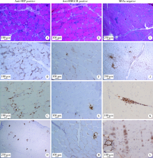
目的 比较不同肌炎特异性自身抗体(myositis specific antibodies,MSAs)类型的免疫介导坏死性肌病(immune-mediated necrotizing myopathies,IMNM)的临床和病理特征。方法 从中日友好医院2008—2018年住院期间所有行肌肉活检的特发性炎性肌病患者中选取符合以下任一条件的IMNM患者104例:(1)抗信号识别颗粒(signal recognition particle,SRP)抗体阳性;(2)抗3-羟基-3-甲基戊二酰辅酶A 还原酶(3-hydroxy-3-methylglutaryl-coenzyme A reductase,HMGCR)抗体阳性;(3)血清MSAs阴性且病理符合2004年欧洲神经肌肉病中心定义的IMNM病理诊断标准。回顾性收集患者的临床、实验室检查和肌肉病理等信息,比较各组临床及病理特征的差异。结果 所有104例IMNM患者中,肌无力(92.3%)、肌酸激酶升高(92.3%)是IMNM的最常见临床表现,此外,33.7%的IMNM患者合并吞咽困难,46.5%的患者合并间质性肺病(interstitial lung diseases,ILD)。抗HMGCR阳性患者较抗SRP阳性和MSAs阴性患者更容易出现V形疹(30.4% vs. 4.3%和5.9%,P<0.01),抗SRP阳性患者合并ILD发生率高于抗HMGCR阳性和MSAs阴性患者(64.4% vs. 34.8%和29.0%,P<0.01),MSAs阴性患者合并其他结缔组织病更多见(32.4% vs. 8.5%和4.3%,P<0.01)。3组IMNM患者肌肉病理中均可见肌细胞坏死(94.2%)、吞噬(65.4%)和再生(67.3%),肌细胞膜表达主要组织相容性复合物-Ⅰ分子上调(78.8%),肌内膜CD4 +T细胞(68.3%)和CD68 +巨噬细胞(65.7%)浸润。结论 抗SRP抗体阳性、抗HMGCR抗体阳性和MSAs阴性的IMNM患者存在异质性,在临床上开展MSAs检测和肌肉病理检查对区分不同类型的IMNM有指导价值。

关键词: 肌炎, 自身免疫病, 信号识别颗粒, 羟甲基戊二酰基CoA还原酶, 病理学

Abstract:

Objective: To investigate the clinical and pathological features of immune-mediated necro-tic myopathies (IMNM) with different myositis-specific antibodies (MSAs).Methods: In the study, 104 IMNM patients who met any of the following three criteria were selected from idiopathic inflammatory myopathy patients who had MSAs results and underwent muscle biopsy from 2008 to 2018 in China-Japan Friendship Hospital: (1) Anti-signal recognition particle (SRP) antibody positive; (2) Anti-3-hydroxy-3-methylglutaryl coenzyme A reductase (HMGCR) antibody positive; (3) MSAs negative and consistent with the pathological diagnostic criteria of IMNM defined by the European Neuromuscular Centre in 2004. The clinical, laboratory and muscle pathological information of the IMNM patients were retrospectively collected and compared in anti-SRP, anti-HMGCR and MSAs negative groups.Results: Of 104 IMNM patients, 47 patients (45.2%) were positive for anti-SRP antibody, 23 (22.1%) were positive for anti-HMGCR antibody, and 34 (32.7%) were negative for MSAs. The common symptoms of IMNM patients were muscle weakness (92.3%), elevated serum creatine kinase level (92.3%), dysphagia (33.7%) and interstitial lung diseases (ILD) (49.5%). The anti-HMGCR-positive patients were more frequent to have “V” sign (30.4% vs. 4.3% and 5.9%, P<0.01) as compared with the anti-SRP-positive and MSAs-negative patients. The incidence of ILD in the anti-SRP-positive patients was higher than that in the anti-HMGCR-positive and MSAs negative patients (64.4% vs. 34.8% and 29.0%, P<0.01). The prevalence of the patients combined with other connective tissue diseases in MSAs-negative IMNM was higher than that in the other two groups (32.4% vs. 8.5% and 4.3%, P<0.01). 93.3% of the anti-SRP-positive patients were found with antinuclear antibody positivity, higher than those of the anti-HMGCR-positive and MSAs-negative patients (93.3% vs. 36.4% and 58.8%, P<0.001). The common pathological features of IMNM were muscle fibre necrosis (94.2%), regeneration (67.3%) and phagocytosis (65.4%), overexpression of major histocompatibility complex-1 on sarcolemma (78.8%), infiltration of CD4 + T cells (81.7%) and CD68 + macrophage (79.8%) and expression of membrane attack complex (MAC) (77.8%). The endomysial infiltration of CD4 + T cells and CD68 + macrophage and MAC expression on sarcolemma in the MSAs-negative group were more common than that in the anti-SRP and anti-HMGCR groups (88.2% vs. 57.4% and 60.9%, 91.2% vs. 59.1% and 38.1%, 76.5% vs. 45.5% and 42.9%, respectively, P<0.01).Conclusion: There is heterogeneity in anti-SRP-positive, anti-HMGCR-positive or MSAs-negative patients. The detection of MSAs and performing of muscle biopsy are useful for distinguishing different types of IMNM.

Key words: Myositis, Autoimmune diseases, Signal recognition particle, Hydroxymethylglutaryl CoA reductases, Pathology

中图分类号: 

  • R593.26

表1

IMNM不同抗体组临床特征的比较"

Features IMNM(n=104) Anti-SRP(n=47) Anti-HMGCR(n=23) MSAs negative(n=34) P
Female 73 (70.2) 38 (80.9) 13 (56.5) 22 (64.7) 0.078
Age/years 46.24±15.38 47.49±15.76 46.09±18.08 44.62±12.02 0.712
Age of onset/years 44.63±16.03 46.11±16.21 44.13±20.83 42.91±11.82 0.671
Duration/months 20.85±27.79 19.01±25.08 25.5±35.03 10.23±26.36 0.652
Chronic progression (>12 months) 59 (56.7) 28 (59.6) 12 (52.2) 19 (55.9) 0.836
Fever 5 (4.8) 3 (6.4) 1 (4.3) 1 (2.9) 0.763
Loss of weight 31 (29.8) 14 (29.8) 4 (17.1) 13 (32.8) 0.241
Muscle weakness 96 (92.3) 44 (93.6) 21 (91.3) 31 (91.1) 0.902
Severe muscle weakness 48 (46.2) 22 (46.8) 9 (39.1) 17 (50.0) 0.716
Muscle atrophy 15/73(20.5) 5/33 (15.6) 4/17 (23.5) 6/24 (25.0) 0.651
Myalgia 29 (27.9) 13 (27.7) 8 (34.8) 8 (23.5) 0.649
Arthritis 10 (9.6) 4 (8.5) 1 (4.3) 5 (14.7) 0.404
Skin involvement 27 (26.0) 12 (25.5) 8 (34.8) 7 (20.6) 0.485
Heliotrope rash 7 (7.7) 3 (6.4) 2 (8.7) 3 (8.8) 0.902
Gottron’s sign 2 (1.9) 1 (2.1) 0 1 (2.9) 0.723
“V” sign 11 (10.6) 2 (4.3) 7 (30.4) 2 (5.9) 0.002*
Shawl sign 6 (5.8) 2 (4.3) 4 (17.4) 0 0.018
Raynaud phenomenon 4 (3.8) 3 (6.4) 0 1 (2.9) 0.404
ILD 46/99 (46.5) 29/45 (64.4) 7/23 (30.4) 10/31 (29.0) 0.004#
Cough 2/46 (4.3) 2/29 (6.9) 0/7 0/10 0.542
Dyspnea 4/46 (8.7) 3/29 (10.3) 0/7 1/10 (10.0) 0.675
Asymptomatic 41/46 (89.1) 25/29 (86.2) 7/7 (100.0) 9/10 (90.0) 0.572
Dysphagia 35 (33.7) 17 (36.2) 8 (34.8) 10 (29.4) 0.576
Cancer 4 (3.8) 3 (6.4) 1 (4.3) 0 0.334
With other CTD 16 (15.4) 4 (8.5) 1 (4.3) 11 (32.4) 0.003
Death 5 (4.8) 3 (6.4) 1 (4.3) 1 (2.9) 0.769

表2

IMNM不同抗体组实验室特征比较"

Features a IMNM (n=104) Anti-SRP (n=47) Anti-HMGCR (n=23) MSAs negative (n=34) P
Anti-Ro-52 25 (24.0) 16 (34.0) 1 (4.3) 8 (23.5) 0.024
ANA 74/101 (73.3) 42/45 (93.3) 8/22 (36.4) 20/34 (58.8) <0.001*
Peak CK/(IU/L) 5 100 (2 749, 9 916) 5 070 (2 975, 9 994) 5 728 (3 320, 11 435) 4 827 (1 324, 9 831) 0.546
ALT (0-40 U/L) 96 (51, 208) 120 (55, 221) 93 (51, 209) 92 (38, 154) 0.500
AST (0-40 U/L) 62 (36, 127) 83 (37, 151) 64 (36, 124) 54 (29, 114) 0.306
LDH (100-250 IU/L) 522 (317, 770) 587 (339, 783) 569 (330, 836) 387 (258, 615) 0.113
CK (26-200 IU/L) 2 157 (810, 4 645) 1 753 (771, 4 241) 2 157 (861, 5 481) 2 326 (612, 4 709) 0.956
CRP (<8 mg/L) 2.9 (1.9, 5.5) 2.9 (1.9, 5.2) 4.3 (2.2, 8.6) 2.6 (1.6, 5.2) 0.198
ESR (<20 mm/h) 8 (4, 16) 9 (4, 20) 8 (4, 20) 6 (3, 13) 0.466
Fet (11.0-306.8 μg/L) 123.0 (48.9, 231.1) 120.0 (58.9, 218.2) 172.0 (109.9, 716.7) 86.0 (22.0, 205.0) 0.126
IgG (6.94-16.20 g/L) 10.50 (8.28, 14.13) 11.60 (8.73, 15.65) 8.59 (6.03, 11.28) 10.50 (8.87, 13.40) 0.033#
IgA (0.68-3.78 g/L) 1.67 (1.17, 2.35) 1.86 (1.35, 2.58) 1.32 (1.03, 1.92) 1.56 (0.09, 2.54) 0.105
IgM (0.60-2.63 g/L) 1.12 (0.79, 1.45) 1.19 (0.82, 1.76) 0.88 (0.66, 1.21) 1.18 (0.84, 1.42) 0.109
C3 (0.70-1.28 g/L) 0.86 (0.75, 1.01) 0.87 (0.77, 0.99) 0.88 (0.78, 1.05) 0.82 (0.71, 1.03) 0.336
C4 (0.16-0.47 g/L) 0.18 (0.15, 0.22) 0.19 (0.15, 0.23) 0.20 (0.17, 0.24) 0.17 (0.12, 0.20) 0.067
T cell (1 000-3 000 cell/mL) 2 060 (1 555, 2 650) 1 870 (1 480, 2 240) 1 990 (1 370, 2 350) 2 620 (1 570, 3 350) 0.027
CD3+ T cell (808-2 072 cell/mL) 1 500 (1 038, 1 877) 1 400 (999, 1 686) 1 458 (903, 1 978) 1 767 (1 125, 2 378) 0.760
CD4+ T cell (380-1 280 cell/mL) 853 (608, 1 283) 817 (604, 1 114) 758 (456, 1 123) 1 767 (1 125, 2 378) 0.042
CD8+ T cell (229-982 cell/μL) 493 (375, 761) 477 (378, 678) 653 (345, 797) 554 (348, 812) 0.586

表3

IMNM不同抗体组间肌肉病理特征比较"

Features IMNM (n=104) Anti-SRP (n=47) Anti-HMGCR (n=23) MSAs negative (n=34) P
Muscle fibre
Necrosis 98 (94.2) 43 (91.5) 21 (91.3) 34 (100.0) 0.213
Regeneration 70 (67.3) 33 (70.2) 11 (47.8) 26 (76.5) 0.066
Phagocytosis 68 (65.4) 27 (57.4) 14 (60.9) 27 (79.4) 0.107
Connective tissue proliferation 36 (34.6) 17 (36.2) 8 (34.8) 11 (32.4) 0.938
MHC-Ⅰ expression on sarcolemma 82 (78.8) 35 (74.5) 17(73.9) 30 (88.2) 0.263
Partial expression 56 (53.8) 24 (51.1) 11 (47.8) 21 (61.8) 0.512
Diffuse expression 26 (25.0) 11 (23.4) 6 (26.1) 9 (26.5) 0.953
Inflammation infiltration
CD4+T cell 85 (81.7) 35 (74.5) 18 (78.3) 32 (94.1) 0.045
Endomysial 71 (68.3) 27 (57.4) 14 (60.9) 30 (88.2) 0.009*
Perivascular 30 (28.8) 17 (36.2) 6 (26.1) 7 (20.6) 0.295
CD8+T cell 57 (54.8) 21 (44.7) 11 (47.8) 25 (73.5) 0.027#
Endomysial 48 (46.2) 17 (36.2) 8 (34.8) 23 (67.6) 0.009*
Perivascular 17 (16.3) 8 (17.0) 4 (17.4) 5 (14.7) 0.951
CD20+B cell 7 (6.7) 2 (4.3) 1 (4.3) 4 (11.8) 0.361
Endomysial 4 (3.8) 1 (2.1) 0 3 (8.8) 0.168
Perivascular 3 (2.9) 1(2.1) 1 (4.3) 1 (2.9) 0.124
CD68+ macrophage 79/99 (79.8) 35/44 (79.5) 11/21 (52.4) 32 (94.1) 0.002
Endomysial 65/99 (65.7) 26/44 (59.1) 8/21 (38.1) 31 (91.2) <0.001*
Perivascular 24/99 (24.2) 18/44 (40.9) 3/21 (15.0) 3 (8.8) 0.003#
MAC 77/99 (77.8) 34/44 (77.3) 11/21 (52.4) 32 (94.1) 0.001
Sarcolemma 55/99 (55.6) 20/44 (45.5) 9/21 (42.9) 26 (76.5) 0.015*
Sarcoplasm 18/99 (18.2) 8/44 (18.2) 3/21 (14.3) 7 (20.6) 0.841
Capillaries 31/99 (31.3) 19/44 (43.2) 4/21 (19.0) 8 (23.5) 0.070

图1

IMNM肌肉病理特征"

[1] Hoogendijk JE, Amato AA, Lecky BR , et al. 119th ENMC international workshop: trial design in adult idiopathic inflammatory myopathies,with the exception of inclusion body myositis[J]. Neuromuscul Disord, 2004,14(5):337-345.
[2] Allenbach Y, Mammen AL, Benveniste O , et al. 224th ENMC International Workshop: clinico-sero-pathological classification of immune-mediated necrotizing myopathies[J]. Neuromuscul Disord, 2018,28(1):87-99.
[3] Pinal-Fernandez I, Mammen AL . Spectrum of immune-mediated necrotizing myopathies and their treatments[J]. Curr Opin Rheumatol, 2016,28(6):619-624.
[4] Lim J, Rietveld A, De Bleecker JL , et al. Seronegative patients form a distinctive subgroup of immune-mediated necrotizing myopathy[J]. Neurol Neuroimmunol Neuroinflamm, 2018,6(1):e513.
[5] Bohan A, Peter JB . Polymyositis and dermatomyositis (first of two parts)[J]. N Engl J Med, 1975,292(7):344-347.
[6] Bohan A, Peter JB . Polymyositis and dermatomyositis (second of two parts)[J]. N Engl J Med, 1975,292(8):403-407.
[7] Suzuki S, Nishikawa A, Kuwana M , et al. Inflammatory myopathy with anti-signal recognition particle antibodies: case series of 100 patients[J]. Orphanet J Rare Dis, 2015,5(13):10-61.
[8] Suzuki S, Hayashi YK, Kuwana M , et al. Myopathy associated with anti- bodies to signal recognition particle: disease progression and neurological outcome[J]. Arch Neurol, 2012,69(6):728-732.
[9] Raghu G, Collard HR, Egan JJ , et al. An official ATSERSJRSALAT statement: idiopathic pulmonary fibrosis: evidence-based guidelines for diagnosis and management[J]. Am J Respir Crit Care Med, 2011,183(6):788-824.
[10] Travis WD, Costabel U, Hansell DM , et al. An official American Thoracic Society European Respiratory Society statement: update of the international multidisciplinary classification of the idiopathic interstitial pneumonias[J]. Am J Respir Crit Care Med, 2013,188(6):733-748.
[11] Dalakas MC . Polymyositis, dermatomyositis and inclusion-body myositis[J]. N Engl J Med, 1991,325(21):1487-1498.
[12] Watanabe Y, Uruha A, Suzuki S , et al. Clinical features and prognosis in anti-SRP and anti-HMGCR necrotising myopathy[J]. Neurol Neurosurg Psychiatry, 2016,87(10):1038-1044.
[13] Takada T, Hirakata M, Suwa A , et al. Clinical and histopatholo-gical features of myopathies in Japanese patients with anti-SRP autoantibodies[J]. Mod Rheumatol, 2009,19(2):156-164.
[14] Ge Y, Lu X, Peng Q , et al. Clinical characteristics of anti-3-hydroxy-3-methylglutaryl coenzyme A reductase antibodies in Chinese patients with idiopathic inflammatory myopathies[J]. PLoS One, 2015,10(10):e0141616.
[15] Limaye V, Bundell C, Hollingsworth P , et al. Clinical and gene-tic associations of autoantibodies to 3-hydroxy-3-methyl-glutaryl-coenzyme a reductase in patients with immune-mediated myositis and necrotizing myopathy[J]. Muscle Nerve, 2015,52(2):196-203.
[16] Christopher-Stine L, Casciola-Rosen LA, Hong G , et al. A novel autoantibody recognizing 200-kD and 100-kD proteins is associated with an immune-mediated necrotizing myopathy[J]. Arthritis Rheum, 2010,62(9):2757-2766.
[17] Mammens AL . Which nonautoimmune myopathies are most frequently misdiagnosed as myositis?[J]. Curr Opin Rheumatol, 2017,29(6):618-622.
[18] Wang Q, Li Y, Ji SQ , et al. Immunopathological characterization of muscle biopsy samples from immune-mediated necrotizing myo-pathy patients[J]. Med Sci Monit, 2018,12(24):2189-2196.
[19] Wang L, Liu LL, Hao HJ , et al. Myopathy with anti-signal recognition particle antibodies: clinical and histopathological features in Chinese patients[J]. Neuromuscular Disorders, 2014,24(4):335-341.
[1] 薛江,张建运,时瑞瑞,谢晓艳,白嘉英,李铁军. 105例口腔颅颌面部纤维性结构不良的临床病理分析[J]. 北京大学学报(医学版), 2022, 54(1): 54-61.
[2] 肖云抒,朱冯赟智,罗澜,邢晓燕,李玉慧,张学武,沈丹华. 88例重叠肌炎的临床及免疫学特征[J]. 北京大学学报(医学版), 2021, 53(6): 1088-1093.
[3] 罗澜,邢晓燕,肖云抒,陈珂彦,朱冯赟智,张学武,李玉慧. 抗合成酶综合征合并心脏受累患者的临床及免疫学特征[J]. 北京大学学报(医学版), 2021, 53(6): 1078-1082.
[4] 张朴丽,杨红霞,张立宁,葛勇鹏,彭清林,王国春,卢昕. 血清YKL-40在诊断抗黑色素瘤分化相关基因5阳性皮肌炎合并严重肺损伤中的价值[J]. 北京大学学报(医学版), 2021, 53(6): 1055-1060.
[5] 伊文霞,魏翠洁,吴晔,包新华,熊晖,常杏芝. 长疗程利妥昔单抗治疗难治性幼年型特发性炎症性肌病3例[J]. 北京大学学报(医学版), 2021, 53(6): 1191-1195.
[6] 吴燕芳,高飞,林滇恬,陈志涵,林禾. 托法替布联合治疗抗MDA5抗体阳性的无肌病皮肌炎并发快速进展型间质性肺病5例临床分析[J]. 北京大学学报(医学版), 2021, 53(5): 1012-1016.
[7] 郑艺明,郝洪军,刘怡琳,郭晶,赵亚雯,张巍,袁云. Ro52抗体与其他肌炎抗体共阳性的相关性研究[J]. 北京大学学报(医学版), 2020, 52(6): 1088-1092.
[8] 甘雨舟,李玉慧,张丽华,马琳,何文雯,金月波,安媛,栗占国,叶华. 临床无肌病性皮肌炎与皮肌炎临床及免疫学特征比较[J]. 北京大学学报(医学版), 2020, 52(6): 1001-1008.
[9] 朱冯赟智,邢晓燕,汤晓菲,李依敏,邵苗,张学武,李玉慧,孙晓麟,何菁. 肌炎合并血栓栓塞患者的临床及免疫学特征[J]. 北京大学学报(医学版), 2020, 52(6): 995-1000.
[10] 刘毅,刘志坚,沈棋,吴静云,范宇,李德润,虞巍,何志嵩. 14例恶性潜能未定的前列腺间质肿瘤病例分析[J]. 北京大学学报(医学版), 2020, 52(4): 621-624.
[11] 郝一昌,颜野,张帆,邱敏,周朗,刘可,卢剑,肖春雷,黄毅,刘承,马潞林. 穿刺活检单针阳性的前列腺癌手术策略选择及经验总结[J]. 北京大学学报(医学版), 2020, 52(4): 625-631.
[12] 马茹,李鑫宝,闫风彩,林育林,李雁. 肿瘤间质比评估阑尾来源腹膜假黏液瘤的临床价值[J]. 北京大学学报(医学版), 2020, 52(2): 240-246.
[13] 张晔,张妮,刘晓筱,周传香. 唾液腺腺样囊性癌淋巴结转移相关的临床病理分析[J]. 北京大学学报(医学版), 2020, 52(1): 30-34.
[14] 付鹏,陈文,崔立刚,葛辉玉,王淑敏. 2017版美国放射学会甲状腺影像学报告与数据系统应用价值探索[J]. 北京大学学报(医学版), 2019, 51(6): 1067-1070.
[15] 徐婧,徐静,李鹤,唐杰,舒建龙,张婧,石连杰,李胜光. 皮肌炎合并IgA血管炎1例[J]. 北京大学学报(医学版), 2019, 51(6): 1173-1177.
Viewed
Full text


Abstract

Cited

  Shared   
  Discussed   
[1] 张三. 中文标题测试[J]. 北京大学学报(医学版), 2010, 42(1): 1 -10 .
[2] 赵磊, 王天龙 . 右心室舒张末期容量监测用于肝移植术中容量管理的临床研究[J]. 北京大学学报(医学版), 2009, 41(2): 188 -191 .
[3] 万有, , 韩济生, John E. Pintar. 孤啡肽基因敲除小鼠电针镇痛作用增强[J]. 北京大学学报(医学版), 2009, 41(3): 376 -379 .
[4] 张燕, 韩志慧, 钟延丰, 王盛兰, 李玲玲, 郑丹枫. 骨骼肌活组织检查病理诊断技术的改进及应用[J]. 北京大学学报(医学版), 2009, 41(4): 459 -462 .
[5] 赵奇, 薛世华, 刘志勇, 吴凌云. 同向施压测定自酸蚀与全酸蚀粘接系统粘接强度[J]. 北京大学学报(医学版), 2010, 42(1): 82 -84 .
[6] 林红, 王玉凤, 吴野平. 学校生活技能教育对小学三年级学生行为问题影响的对照研究[J]. 北京大学学报(医学版), 2007, 39(3): 319 -322 .
[7] 丰雷, 程嘉, 王玉凤. 注意缺陷多动障碍儿童的运动协调功能[J]. 北京大学学报(医学版), 2007, 39(3): 333 -336 .
[8] 李岳玲, 钱秋瑾, 王玉凤. 儿童注意缺陷多动障碍成人期预后及其预测因素[J]. 北京大学学报(医学版), 2007, 39(3): 337 -340 .
[9] . 书讯[J]. 北京大学学报(医学版), 2007, 39(3): 225 -328 .
[10] 牟向东, 王广发, 刁小莉, 阙呈立. 肺黏膜相关淋巴组织型边缘区B细胞淋巴瘤一例[J]. 北京大学学报(医学版), 2007, 39(4): 346 -350 .