北京大学学报(医学版) ›› 2021, Vol. 53 ›› Issue (2): 255-260. doi: 10.19723/j.issn.1671-167X.2021.02.004
血浆Dickkopf-1在类风湿关节炎患者中的表达及其与外周血T细胞亚群的相关性
曹迪1,王燕1,Δ( ),王柳青1,孙晓麟2,黄妃3,孟洋1,任丽丽1,张学武2,Δ()
- 1.郑州大学第五附属医院风湿免疫科, 郑州 450000
2.北京大学人民医院风湿免疫科, 北京 100044
3.遵义医科大学附属医院肾病风湿科, 贵州遵义 563000
Expression of plasma Dickkopf-1 in patients with rheumatoid arthritis and its correlation with peripheral blood T cell subsets
CAO Di1,WANG Yan1,Δ(),WANG Liu-qing1,SUN Xiao-lin2,HUANG Fei3,MENG Yang1,REN Li-li1,ZHANG Xue-wu2,Δ()
- 1. Department of Rheumatology and Immunology, The Fifth Affiliated Hospital of Zhengzhou University, Zhengzhou 450000, China
2. Department of Rheumatology and Immunology, Peking University People’s Hospital, Beijing 100044, China
3. Department of Rheumatology and Immunology,Affiliated Hospital of Zunyi Medical University, Zunyi 563000, Guizhou, China
摘要:
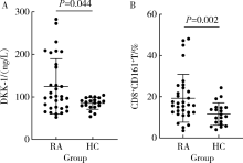
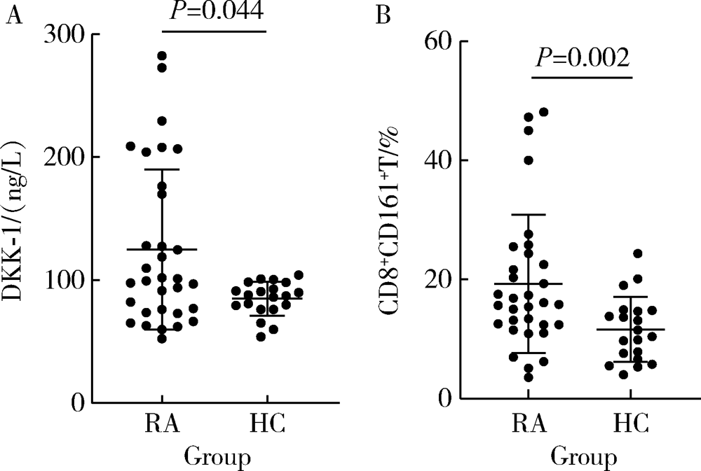
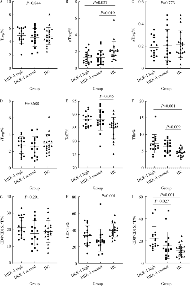
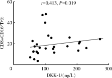
目的: 检测类风湿关节炎(rheumatoid arthritis,RA)患者血浆Dickkopf-1(DKK-1)水平,并分析其与外周血T细胞亚群、临床指标之间的相关性。方法: 采用酶联免疫吸附法(enzyme linked immunosorbent assay,ELISA)检测32例RA患者及20例健康对照者的血浆DKK-1的水平,并记录RA患者的各项临床表现和实验室指标。流式细胞仪检测RA患者外周血T细胞亚群(包括Treg、nTreg、aTreg、sTreg、Teff、Tfh、CD4+CD161+T、CD8+T、CD8+CD161+T细胞)。比较两组血浆DKK-1的水平,并分析其与外周血T细胞亚群、临床指标的相关性。结果: (1)RA患者血浆DKK-1浓度为(124.97±64.98) ng/L,健康对照组血浆DKK-1浓度为(84.95±13.74) ng/L,RA患者血浆DKK-1水平显著高于健康对照组(P<0.05),RA患者外周血CD8+CD161+T细胞百分比显著高于健康对照组(P<0.05)。(2)血浆DKK-1水平与红细胞沉降率(r=0.406,P=0.021)、DAS28评分(r=0.372,P=0.036)、免疫球蛋白G(r=0.362,P=0.042)、免疫球蛋白A(r=0.377,P=0.033)呈正相关;与年龄、病程、C-反应蛋白、类风湿因子、抗环瓜氨酸肽抗体、免疫球蛋白M、补体C3、补体C4、白细胞计数、中性粒细胞百分比无相关性。(3)RA患者血浆DKK-1水平与外周血CD8+CD161+T细胞百分比呈正相关(r=0.413,P=0.019);与Treg、nTreg、aTreg、sTreg、Teff、Tfh、CD4+CD161+T、CD8+T细胞无相关性。(4)外周血CD161+CD8+T细胞百分比与红细胞沉降率(r=-0.415,P=0.004)、C-反应蛋白(r=-0.393,P=0.007)、DAS28评分(r=-0.392,P=0.007)、类风湿因子(r=-0.535,P<0.001)、抗环瓜氨酸肽抗体(r=-0.589,P<0.001)、免疫球蛋白G(r=-0.368,P=0.012)、免疫球蛋白M(r=-0.311,P=0.035)呈负相关;与年龄、病程、免疫球蛋白A、补体C3、补体C4、白细胞计数、中性粒细胞百分比无相关性。结论: RA患者血浆DKK-1水平和外周血T细胞亚群中的CD8+CD161+T细胞百分比均升高,可能与患者体内促炎细胞因子的分泌有关;DKK-1参与骨稳态的调节,可用作RA骨破坏的标志物。
中图分类号:
- R593.2



| [1] |
Smolen JS, Aletaha D, Mcinnes IB. Rheumatoid arthritis[J]. Lancet, 2016,388(10055):2023-2038.
pmid: 27156434 |
| [2] | Lee GR. The balance of Th17 versus Treg cells in autoimmunity[J]. Int J Mol Sci, 2018,19(3):730. |
| [3] |
Chae WJ, Park JH, Henegariu O, et al. Membrane-bound Dickkopf-1 in Foxp3+ regulatory T cells suppresses T-cell-mediated autoimmune colitis[J]. Immunology, 2017,152(2):265-275.
pmid: 28556921 |
| [4] |
Chae WJ, Ehrlich AK, Chan PY, et al. The Wnt antagonist Dickkopf-1 promotes pathological type 2 cell-mediated Inflammation[J]. Immunity, 2016,44(2):246-258.
pmid: 26872695 |
| [5] | Aletaha D, Neogi T, Silman AJ, et al. 2010 rheumatoid arthritis classification criteria: an American College of Rheumatology/European League Against Rheumatism collaborative initiative[J]. Ann Rheum Dis, 2010,62(9):2569-2581. |
| [6] |
Glinka A, Wu W, Delius H, et al. Dickkopf-1 is a member of a new family of secreted proteins and functions in head induction[J]. Nature, 1998,391(6665):357-362.
pmid: 9450748 |
| [7] | Fujita K, Janz S. Attenuation of WNT signaling by DKK-1 and -2 regulates BMP2-induced osteoblast differentiation and expression of OPG, RANKL and M-CSF[J]. Mol Cancer, 2007,6(1):71. |
| [8] |
Diarra D, Stolina M, Polzer K, et al. Dickkopf-1 is a master regulator of joint remodeling[J]. Nat Med, 2007,13(2):156-163.
pmid: 17237793 |
| [9] | Gómez-Vaquero C, Martín I, Loza E, et al. Effect of osteoprotegerin and Dickkopf-related protein 1 on radiological progression in tightly controlled rheumatoid arthritis[J]. PLoS One, 2016,11(12):e0166691. |
| [10] |
Rout N. Enhanced Th1/Th17 functions of CD161+CD8+T cells in mucosal tissues of rhesus macaques[J]. PLoS One, 2016,11(6):e0157407.
pmid: 27309719 |
| [11] | Mitsuo A, Morimoto S, Nakiri Y, et al. Decreased CD161+CD8+ T cells in the peripheral blood of patients suffering from rheumatic diseases[J]. Rheumatology (Oxford), 2006,45(12):1477-1484. |
| [12] |
Nicol B, Salou M, Vogel I, et al. An intermediate level of CD161 expression defines a novel activated, inflammatory, and pathogenic subset of CD8+T cells involved in multiple sclerosis[J]. J Autoimmun, 2018,88:61-74.
doi: 10.1016/j.jaut.2017.10.005 |
| [1] | 魏慧, 张警丰, 姚中强, 赵金霞. 类风湿关节炎合并慢性病贫血患者的临床特征及相关因素[J]. 北京大学学报(医学版), 2026, 58(2): 307-312. |
| [2] | 练益瑞, 刘静璇, 赵亮, 赵静, 臧思田, 李玉慧. 抗PM/Scl抗体相关风湿性疾病谱及其在特发性炎性肌病中的免疫学特征[J]. 北京大学学报(医学版), 2025, 57(6): 1018-1023. |
| [3] | 吴滔, 林建子, 朱亚锋, 马剑达, 贾霈雯, 杨莉娟, 潘婕, 邹耀威, 杨迎, 卢烨, 戴冽. 血清蛋白质谱筛选及验证类风湿关节炎患者肌肉量减少的生物标志物[J]. 北京大学学报(医学版), 2025, 57(6): 1024-1031. |
| [4] | 丁艳, 王丽芳, 李超然, 卢哲敏, 石连杰. 利妥昔单抗成功治疗类风湿关节炎合并IgG4相关性疾病1例[J]. 北京大学学报(医学版), 2025, 57(6): 1203-1207. |
| [5] | 杨菊, 徐婧, 戴菊华, 石连杰. Lumican蛋白在类风湿关节炎患者血清中的表达及其与疾病和免疫活动的相关性[J]. 北京大学学报(医学版), 2025, 57(5): 911-918. |
| [6] | 冯亮华, 洪丽荣, 陈雨佳, 蔡学明. 泛素特异性蛋白酶35对类风湿关节炎成纤维样滑膜细胞铁死亡的作用及机制[J]. 北京大学学报(医学版), 2025, 57(5): 919-925. |
| [7] | 贾霈雯, 杨迎, 邹耀威, 欧阳志明, 林建子, 马剑达, 杨葵敏, 戴冽. 类风湿关节炎患者低肌肉量综合征的临床特征及其对躯体功能的影响[J]. 北京大学学报(医学版), 2024, 56(6): 1009-1016. |
| [8] | 马豆豆, 卢哲敏, 郭倩, 朱莎, 古今, 丁艳, 石连杰. 小剂量利妥昔单抗成功治疗类风湿关节炎合并重症肌无力1例[J]. 北京大学学报(医学版), 2024, 56(6): 1110-1114. |
| [9] | 王明霞, 丁菱, 王敏, 邹婵娟, 颜丝语, 梁颖文, 王伟佳, 何善智. 双靶点嵌合抗原受体T细胞治疗系统性红斑狼疮患者停药后安全孕产1例[J]. 北京大学学报(医学版), 2024, 56(6): 1119-1125. |
| [10] | 闫蕊, 柯丹, 张妍, 李丽, 苏焕然, 陈伟, 孙明霞, 刘晓敏, 罗靓. 血清趋化因子CXCL-10和涎液化糖链抗原6水平在类风湿关节炎合并肺间质病变患者中的诊断和病情评估价值[J]. 北京大学学报(医学版), 2024, 56(6): 956-962. |
| [11] | 赵亮, 史成龙, 马柯, 赵静, 王潇, 邢晓燕, 莫万星, 练益瑞, 高超, 李玉慧. 抗合成酶综合征重叠类风湿关节炎患者的免疫学特征[J]. 北京大学学报(医学版), 2024, 56(6): 972-979. |
| [12] | 韩艺钧, 陈小莉, 李常虹, 赵金霞. 甲氨蝶呤在类风湿关节炎患者中的应用现状[J]. 北京大学学报(医学版), 2024, 56(6): 994-1000. |
| [13] | 刘东武, 陈杰, 高明利, 于静. 类风湿关节炎伴发淋巴结Castleman样病理改变1例[J]. 北京大学学报(医学版), 2024, 56(5): 928-931. |
| [14] | 黄会娜,赵静,赵祥格,白自然,李霞,王冠. 乳酸对类风湿关节炎患者外周血CD4+T细胞亚群的调控作用[J]. 北京大学学报(医学版), 2024, 56(3): 519-525. |
| [15] | 汤晓菲,李永红,丁秋玲,孙卓,张阳,王育梅,田美伊,刘坚. 类风湿关节炎患者下肢深静脉血栓发病率及危险因素[J]. 北京大学学报(医学版), 2024, 56(2): 279-283. |
|
||
