北京大学学报(医学版) ›› 2021, Vol. 53 ›› Issue (3): 598-601. doi: 10.19723/j.issn.1671-167X.2021.03.027
碘液浸染在Micro-CT下识别小鼠颅底-颞下区肿瘤组织中的应用
杨榕1,李庆祥1,王逸飞1,周闻2,王雯3,郭传瑸1,刘浩2,Δ( ),郭玉兴1,Δ()
- 1.北京大学口腔医学院·口腔医院,口腔颌面外科 国家口腔疾病临床医学研究中心 口腔数字化医疗技术和材料国家工程实验室 口腔数字医学北京市重点实验室,北京 100081
2.北京大学口腔医学院·口腔医院中心实验室,北京 100081
3.河北医科大学口腔医院正畸科,石家庄 050017
Application of iodine staining technique for tumor identification in Micro-CT of mouse model with skull base-infratemporal fossa tumor
YANG Rong1,LI Qing-xiang1,WANG Yi-fei1,ZHOU Wen2,WANG Wen3,GUO Chuan-bin1,LIU Hao2,Δ(),GUO Yu-xing1,Δ()
- 1. Department of Oral and Maxillofacial Surgery,Peking University School and Hospital of Stomatology & National Clinical Research Center for Oral Diseases & National Engineering Laboratory for Digital and Material Technology of Stomatology & Beijing Key Laboratory of Digital Stomatology, Beijing 100081, China
2. Central Laboratory,Peking University School and Hospital of Stomatology, Beijing 100081, China
3. Department of Orthodontics, Hospital of Stomotology, Hebei Medical University, Shijiazhuang 050017, China
摘要:
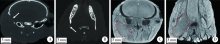
目的: 建立颅底-颞下区恶性肿瘤动物模型,探索碘液浸染技术在Micro-CT图像中识别肿瘤组织的作用。方法: 对12只BABL/c裸鼠采用异氟烷吸入镇静麻醉,在小动物超声系统引导下经颌下区注射头颈鳞状细胞癌WSU-HN6细胞至右侧颞下窝。观察3周后解剖头颅标本,用4%多聚甲醛固定,并行Micro-CT扫描,3.75%复方碘液浸染后重复扫描。将头部标本包埋、切片,进行苏木精-伊红染色和免疫组织化学染色分析肿瘤形成情况。结果: 经Micro-CT分析发现颅骨有明显破坏,但无法辨别肿瘤组织;经3.75%复方碘液浸染后,在Micro-CT阅读软件中可以清晰观察肿瘤及周围软组织形态。经苏木精-伊红染色和免疫组织化学染色分析证实,颅底-颞下区形成鳞状细胞癌,同时伴有明显颅骨破坏。结论: 采用颌下注射方式可以成功构建颅底-颞下区肿瘤动物模型;Micro-CT可以观察到颅骨骨质改变,采用复方碘液浸染后有利于观察肿瘤及周围软组织结构。
中图分类号:
- R329.4



| [1] | 阮彩莲, 杨延庆, 薛涛, 等. 经耳前颞叶底入路显露中颅底和岩斜区的便携式视频显微解剖[J]. 解剖学报, 2016,47(4):507-509. |
| [2] |
Guo Y, Guo C. Maxillary-fronto-temporal approach for removal of recurrent malignant infratemporal fossa tumors: Anatomical and clinical study[J]. J Craniomaxillofac Surg, 2014,42(3):206-212.
doi: 10.1016/j.jcms.2013.05.001 |
| [3] | 杨榕, 李庆祥, 毛驰, 等. 多模态影像融合技术与颅底-颞下区肿瘤的诊断和治疗[J]. 北京大学学报(医学版), 2019,51(1):53-58. |
| [4] | 郭玉兴, 彭歆, 刘筱菁, 等. 导航技术在颅底-颞下区肿瘤手术中的应用[J]. 中华口腔医学杂志, 2013,48(11):645-647. |
| [5] | 郭玉兴, 郭传瑸. 增强CT三维重建在颞下咽旁间隙肿瘤中的应用[J]. 北京大学学报(医学版), 2011,43(1):148-150. |
| [6] | 郭玉兴, 郭传瑸, 俞光岩, 等. 影响颞下咽旁间隙恶性肿瘤预后的因素分析[J]. 中华神经外科杂志, 2012,28(8):775-779. |
| [7] | 毛以华, 朱昭炜, 丁茂超, 等. 应用高分辨率显微CT进行大鼠周围神经微血管三维可视化研究[J]. 解剖学报, 2013,44(3):353-356. |
| [8] |
Faraj KA, Cuijpers VM, Wismans RG, et al. Micro-computed tomographical imaging of soft biological materials using contrast techniques[J]. Tissue Eng Part C Methods, 2009,15(3):493-499.
doi: 10.1089/ten.tec.2008.0436 |
| [9] |
Degenhardt K, Wright AC, Horng D, et al. Rapid 3D phenotyping of cardiovascular development in mouse embryos by micro-CT with iodine staining[J]. Circ Cardiovasc Imaging, 2010,3(3):314-322.
doi: 10.1161/CIRCIMAGING.109.918482 pmid: 20190279 |
| [10] |
Jeffery NS, Stephenson RS, Gallagher JA, et al. Micro-computed tomography with iodine staining resolves the arrangement of muscle fibres[J]. J Biomech, 2011,44(1):189-192.
doi: 10.1016/j.jbiomech.2010.08.027 pmid: 20846653 |
| [11] |
Metscher BD. MicroCT for developmental biology: a versatile tool for high-contrast 3D imaging at histological resolutions[J]. Dev Dyn, 2009,238(3):632-640.
doi: 10.1002/dvdy.v238:3 |
| [12] |
Wu J, Yin N. Anatomy research of nasolabial muscle structure in fetus with cleft lip: an iodine staining technique based on microcomputed tomography[J]. J Craniofac Surg, 2014,25(3):1056-1061.
doi: 10.1097/SCS.0000000000000651 |
| [13] |
Wu J, Yin N. Detailed anatomy of the nasolabial muscle in human fetuses as determined by Micro-CT combined with iodine staining[J]. Ann Plast Surg, 2016,76(1):111-116.
doi: 10.1097/SAP.0000000000000219 |
| [14] | 崔国峰, 魏戎, 武军龙, 等. 骨关节炎动物模型的综合评估[J]. 中华骨与关节外科杂志, 2019,12(1):68-74. |
| [1] | 季加孚, 韦静涛, 季科, 步召德. 胃癌诊疗的瓶颈与破局:迈向精准化与智能化融合的新纪元[J]. 北京大学学报(医学版), 2026, 58(2): 231-238. |
| [2] | 高加勒, 张忠涛. 局部进展期直肠癌精准治疗现状与展望[J]. 北京大学学报(医学版), 2026, 58(2): 247-250. |
| [3] | 王海, 江一舟. 靶向血管治疗在乳腺癌精准治疗中的分子机制与临床应用[J]. 北京大学学报(医学版), 2026, 58(2): 251-256. |
| [4] | 罗必显, 刘洪铭, 谢伟勋, 龚渭华. 产甲胎蛋白胃癌的新临床特征和前沿科学问题[J]. 北京大学学报(医学版), 2026, 58(2): 257-265. |
| [5] | 杜文, 章文博, 于尧, 刘硕, 苏惠裕, 胡耒豪, 唐祖南, 吴彬彰, 陈震, 李家琦, 王昊, 彭歆. 口腔颌面部肿瘤"数智化外科"诊疗流程探索与临床应用[J]. 北京大学学报(医学版), 2026, 58(2): 278-284. |
| [6] | 王楠楠, 袁大晋, 朱昱冰, 丁磊. 结直肠癌根治术后肝转移风险多中心列线图预测模型的构建与验证[J]. 北京大学学报(医学版), 2026, 58(2): 290-300. |
| [7] | 刘友东, 吕亚军, 陈杰, 臧明德, 潘宏达, 刘晓文, 陆俊, 刘凤林. 全腹腔镜保留贲门胃底胃次全切除术治疗中上部胃癌的疗效及安全性[J]. 北京大学学报(医学版), 2026, 58(2): 301-306. |
| [8] | 李嘉临, 陈力侨, 唐家天, 吴艳, 王安强. 胃肝样腺癌转化治疗1例[J]. 北京大学学报(医学版), 2026, 58(2): 399-404. |
| [9] | 李斌, 梁寒. 机器人胃癌根治术:研究进展与实践挑战[J]. 北京大学学报(医学版), 2026, 58(2): 416-422. |
| [10] | 董海峰, 陈恒星, 张常华. 恶性肿瘤中蛋白质乳酸化修饰的研究进展[J]. 北京大学学报(医学版), 2026, 58(2): 423-430. |
| [11] | 李宏杨, 黄涛, 王琳琳. 脂肪肌肉比率与卵巢良性肿瘤风险的关联性[J]. 北京大学学报(医学版), 2026, 58(1): 169-174. |
| [12] | 高若凡, 马天宇, 王润楷, 殷雨辰, 李芮迪, 王丹丹, 夏斌. 细胞膜囊泡递送靶向肿瘤坏死因子-α的小干扰RNA对牙髓干细胞的抗炎作用[J]. 北京大学学报(医学版), 2026, 58(1): 22-29. |
| [13] | 刘艳华, 陆敏, 赵旭阳, 张宽根, 武睿, 梅放, 戴志豪, 由江峰, 裴斐. 肿瘤转移抑制基因LASS2去磷酸化对液泡型ATP酶活性及前列腺癌侵袭性的影响[J]. 北京大学学报(医学版), 2025, 57(6): 1113-1123. |
| [14] | 杨小勇, 张帆, 马潞林, 刘承. 前列腺导管腺癌临床特征及腺外侵犯的影响因素[J]. 北京大学学报(医学版), 2025, 57(5): 956-960. |
| [15] | 陈定一, 杜浩鑫, 张逸晨, 王闫飞, 刘巍, 焦园园, 史录文, 管晓东, 卢新璞. 姑息治疗对晚期癌症患者药物使用和医疗资源利用的影响[J]. 北京大学学报(医学版), 2025, 57(5): 996-1001. |
|
||
