北京大学学报(医学版) ›› 2021, Vol. 53 ›› Issue (5): 983-989. doi: 10.19723/j.issn.1671-167X.2021.05.029
下颌骨颏部骨折联合双侧髁突囊内骨折致伤机制的三维有限元分析
周伟1,安金刚1,△( ),荣起国2,△(),张益1
- 1.北京大学口腔医学院·口腔医院,口腔颌面外科 国家口腔医学中心 国家口腔疾病临床医学研究中心 口腔数字化医疗技术和材料国家工程实验室,北京 100081
2.北京大学工学院力学与工程科学系,北京 100871
Three-dimensional finite element analysis of traumatic mechanism of mandibular symphyseal fracture combined with bilateral intracapsular condylar fractures
ZHOU Wei1,AN Jin-gang1,△(),RONG Qi-guo2,△(),ZHANG Yi1
- 1. Department of Oral and Maxillofacial Surgery, Peking University School and Hospital of Stomatology & National Center of Stomatology & National Clinical Research Center for Oral Diseases & National Engineering Laboratory for Digital and Material Technology of Stomatology, Beijing 100081, China
2. Department of Mechanics and Engineering Science, College of Engineering, Peking University, Beijing 100871, China
摘要:
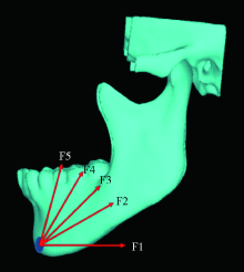
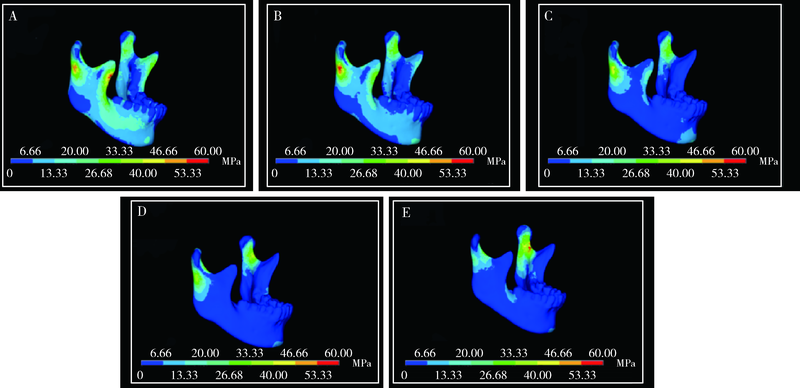
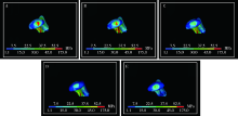
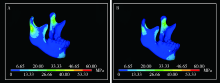
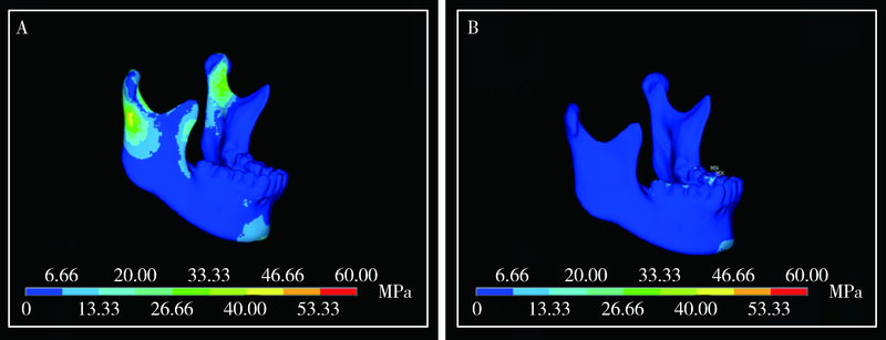
目的: 通过三维有限元方法模拟和分析下颌骨颏部骨折联合双侧髁突囊内骨折的致伤机制,提高此类骨折的预防和诊断水平。方法: 获取1名下颌骨发育正常、无第三磨牙、无颞下颌关节病史的青年男性的颌面部CT和颞下颌关节MRI数据,通过Mimics和ANSYS软件建立三维有限元模型。采取不同角度的外力作用于下颌骨颏部,分析下颌骨及髁突关节面的应力分布。同时,比较有无关节盘、咬合与非咬合状态下下颌骨的应力分布差异。结果: 准确建立了包含颞下颌关节结构的三维有限元模型。当外力作用于下颌骨颏部时,髁突、升支前缘及颏部受力区是主要的应力集中区,其中应力最大值位于髁突顶部。随着外力方向与水平面夹角由0°逐渐增大直至60°,下颌骨上的应力由分散逐渐集中至颏部与双侧髁突三个部位;超过60°时,应力又出现分散的趋势。当对关节盘进行模拟后,髁突关节面及髁颈部的应力分布明显减小。与非咬合状态相比,咬合状态下下颌骨上的应力集中于咬合面,而其他部位无明显的应力分布。结论: 外力方向与水平面呈60°时,应力分布主要集中于颏部及双侧髁突顶部,即三点骨折发生的部位;在下颌骨颏部受力过程中,关节盘的存在以及稳定的咬合状态下,髁突部位(包括颈部和关节面)的应力分布明显减小。
中图分类号:
- R782.6


| [1] | 武付花, 黄迪炎, 郭振国, 等. 三维有限元分析下颌骨不同部位受力髁突的力学应变 [J]. 中国组织工程研究, 2015, 19(29):4667-4671. |
| [2] |
Gallas-Torreira M, Fernandez JR. A three-dimensional computer model of the human mandible in two simulated standard trauma situations [J]. J Craniomaxillofac Surg, 2004, 32(5):303-307.
doi: 10.1016/j.jcms.2004.04.008 |
| [3] | 徐晓峰, 苏佳楠, 代杰文, 等. 颏部骨折合并双侧髁突囊内骨折3种不同治疗方式的三维有限元分析 [J]. 中国口腔颌面外科杂志, 2016, 14(5):409-412. |
| [4] |
Liu YF, Wang R, Baur DA, et al. A finite element analysis of the stress distribution to the mandible from impact forces with various orientations of third molars [J]. J Zhejiang Univ Sci B, 2018, 19(1):38-48.
doi: 10.1631/jzus.B1600552 |
| [5] |
Loukota RA, Neff A, Rasse M. Nomenclature/classification of fractures of the mandibular condylar head [J]. Br J Oral Maxillofac Surg, 2010, 48(6):477-478.
doi: 10.1016/j.bjoms.2009.08.036 pmid: 19896755 |
| [6] |
Antic S, Vukicevic AM, Milasinovic M, et al. Impact of the lower third molar presence and position on the fragility of mandibular angle and condyle: A Three-dimensional finite element study [J]. J Craniomaxillofac Surg, 2015, 43(6):870-878.
doi: 10.1016/j.jcms.2015.03.025 |
| [7] |
Thresher RW, Saito GE. The stress analysis of human teeth [J]. J Biomech, 1973, 6(5):443-449.
pmid: 4748494 |
| [8] |
Arne W, Wolfgang K, Kurt S, et al. A 3-dimensional finite-element analysis investigating the biomechanical behavior of the mandible and plate osteosynjournal in cases of fractures of the condylar process [J]. Oral Surg Oral Med Oral Pathol Oral Radiol Endod, 2002, 94(6):678-686.
doi: 10.1067/moe.2002.126451 |
| [9] |
Rho JY, Hobatho MC, Ashman RB. Relations of mechanical properties to density and CT numbers in human bone [J]. Med Eng Phys, 1995, 17(5):347-355.
pmid: 7670694 |
| [10] |
Wang R, Liu Y, Wang JH, et al. Effect of interfragmentary gap on the mechanical behavior of mandibular angle fracture with three fixation designs: A finite element analysis [J]. J Plast Reconstr Aesthet Surg, 2017, 70(3):360-369.
doi: 10.1016/j.bjps.2016.10.026 |
| [11] |
Chaudhry H, Bukiet B, Anderson EZ, et al. Muscle strength and stiffness in resistance exercise: Force transmission in tissues [J]. J Bodyw Mov Ther, 2017, 21(3):517-522.
doi: S1360-8592(16)30128-0 pmid: 28750958 |
| [12] |
Weijs WA, Hillen B. Relationship between the physiological cross-section of the human jaw muscles and their cross-sectional area in computer tomograms [J]. Acta Anat, 1984, 121(1):31-35.
doi: 10.1159/000145938 |
| [13] |
Bezerra TP, Silva Junior FI, Scarparo HC, et al. Do erupted third molars weaken the mandibular angle after trauma to the chin region? A 3D finite element study [J]. Int J Oral Maxillofac Surg, 2013, 42(4):474-480.
doi: 10.1016/j.ijom.2012.10.009 |
| [14] |
Santos LS, Rossi AC, Freire AR, et al. Finite-element analysis of 3 situations of trauma in the human edentulous mandible [J]. J Oral Maxillofac Surg, 2015, 73(4):683-691.
doi: 10.1016/j.joms.2014.10.014 |
| [15] |
Stringhini DJ, Sommerfeld R, Uetanabaro LC, et al. Resistance and stress finite element analysis of different types of fixation for mandibular orthognathic surgery [J]. Braz Dent J, 2016, 27(3):284-291.
doi: 10.1590/0103-6440201600336 pmid: 27224561 |
| [16] |
Bujtar P, Sandor GK, Bojtos A, et al. Finite element analysis of the human mandible at 3 different stages of life [J]. Oral Surg Oral Med Oral Pathol Oral Radiol Endod, 2010, 110(3):301-309.
doi: 10.1016/j.tripleo.2010.01.025 |
| [17] | Sun M, Yang J, Zhou R, et al. Mechanical analysis on individualized finite element of temporal-mandibular joint under overlarge jaw opening status [J]. Int J Clin Exp Med, 2015, 8(6):9046. |
| [18] |
Cheng HY, Peng PW, Lin YJ, et al. Stress analysis during jaw movement based on vivo computed tomography images from patients with temporomandibular disorders [J]. Int J Oral Maxillofac Surg, 2013, 42(3):386-392.
doi: 10.1016/j.ijom.2012.07.005 |
| [19] |
Chen S, Zhang Y, An JG, et al. Width-controlling fixation of symphyseal/parasymphyseal fractures associated with bilateral condylar fractures with 22.0 mm miniplates: A retrospective investigation of 45 cases [J]. J Oral Maxillofac Surg, 2016, 74(2):315-327.
doi: 10.1016/j.joms.2015.09.030 |
| [1] | 王昕莹, 程雪原, 张孟钧, 李菲, 段晋瑜, 乔静. 浓缩生长因子联合引导性组织再生术治疗下颌磨牙根分叉病变的疗效[J]. 北京大学学报(医学版), 2026, 58(2): 372-379. |
| [2] | 仇师禹, 练洋, 康一帆, 张雷, 蔡义望, 单小峰, 蔡志刚. 基于下颌骨数据库和全连接神经网络的三维检索模型辅助下的下颌骨个性化重建[J]. 北京大学学报(医学版), 2025, 57(2): 360-368. |
| [3] | 马丽娟, 腾雍辉, 王勇, 赵一姣, 张馨月, 秦庆钊, 尹东. 乳牙缺失数字化丝圈间隙保持器的三维有限元分析[J]. 北京大学学报(医学版), 2025, 57(2): 376-383. |
| [4] | 王菲, 张馨月, 刘木清, 王恩博, 段登辉. 顺牙长轴拔牙法在下颌近中与水平智齿拔除术中的应用及三维有限元分析[J]. 北京大学学报(医学版), 2025, 57(1): 106-112. |
| [5] | 康一帆, 葛严军, 吕晓鸣, 谢尚, 单小峰, 蔡志刚. 即刻种植体支持式义齿修复的血管化髂骨瓣重建下颌骨缺损[J]. 北京大学学报(医学版), 2025, 57(1): 78-84. |
| [6] | 胡轶博, 吕伟佳, 夏炜, 刘亦洪. 基于细胞生长与成骨分化的不同孔径生物支架流体力学有限元分析[J]. 北京大学学报(医学版), 2025, 57(1): 97-105. |
| [7] | 徐心雨,吴灵,宋凤岐,李自力,张益,刘筱菁. 基于下颌运动轨迹的正颌外科术中下颌骨髁突定位方法及初步精度验证[J]. 北京大学学报(医学版), 2024, 56(1): 57-65. |
| [8] | 王聪伟,高敏,于尧,章文博,彭歆. 游离腓骨瓣修复下颌骨缺损术后义齿修复的临床分析[J]. 北京大学学报(医学版), 2024, 56(1): 66-73. |
| [9] | 卢汉,张建运,杨榕,徐乐,李庆祥,郭玉兴,郭传瑸. 下颌牙龈鳞状细胞癌患者预后的影响因素[J]. 北京大学学报(医学版), 2023, 55(4): 702-707. |
| [10] | 欧蒙恩,丁云,唐卫峰,周永胜. 基台边缘-牙冠的平台转移结构中粘接剂流动的三维有限元分析[J]. 北京大学学报(医学版), 2023, 55(3): 548-552. |
| [11] | 高梓翔,王勇,温奥楠,朱玉佳,秦庆钊,张昀,王晶,赵一姣. 基于三维下颌骨平均模型的颌骨标志点自动确定方法[J]. 北京大学学报(医学版), 2023, 55(1): 174-180. |
| [12] | 马珂楠,陈虎,沈妍汝,周永胜,王勇,孙玉春. 选择性激光熔化打印可摘局部义齿圆环形卡环固位力的有限元分析[J]. 北京大学学报(医学版), 2022, 54(1): 105-112. |
| [13] | 任爽,时会娟,张家豪,刘振龙,邵嘉艺,朱敬先,胡晓青,黄红拾,敖英芳. 前交叉韧带重建术后移植物应力的有限元分析[J]. 北京大学学报(医学版), 2021, 53(5): 865-870. |
| [14] | 姜又升,冯琳,高学军. 垫底材料弹性模量对髓腔固位冠修复后上颌前磨牙应力分布的影响[J]. 北京大学学报(医学版), 2021, 53(4): 764-769. |
| [15] | 林春平,卢松鹤,朱浚鑫,胡洪成,岳兆国,唐志辉. 个性化根形种植体的螺纹形态对周围牙槽骨应力分布影响的三维有限元分析[J]. 北京大学学报(医学版), 2019, 51(6): 1130-1137. |
|
||
