北京大学学报(医学版) ›› 2023, Vol. 55 ›› Issue (2): 243-253. doi: 10.19723/j.issn.1671-167X.2023.02.007

• 论著 • 上一篇    下一篇

人表皮生长因子受体2低表达乳腺癌的临床病理学特征及预后

朱晓娟1,张虹1,*(),张爽1,李东1,李鑫1,徐玲2,李挺1   

  1. 1. 北京大学第一医院病理科,北京 100034
    2. 北京大学第一医院乳腺疾病中心,北京 100034
  • 收稿日期:2022-11-30 出版日期:2023-04-18 发布日期:2023-04-12
  • 通讯作者: 张虹 E-mail:zhanghong-path@pkufh.com

Clinicopathological features and prognosis of breast cancer with human epidermal growth factor receptor 2 low expression

Xiao-juan ZHU1,Hong ZHANG1,*(),Shuang ZHANG1,Dong LI1,Xin LI1,Ling XU2,Ting LI1   

  1. 1. Department of Pathology, Peking University First Hospital, Beijing 100034, China
    2. Breast Disease Center, Peking University First Hospital, Beijing 100034, China
  • Received:2022-11-30 Online:2023-04-18 Published:2023-04-12
  • Contact: Hong ZHANG E-mail:zhanghong-path@pkufh.com

摘要:

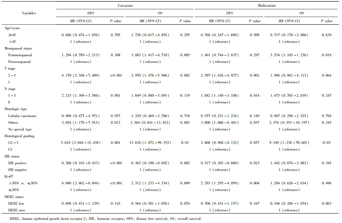
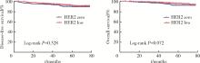
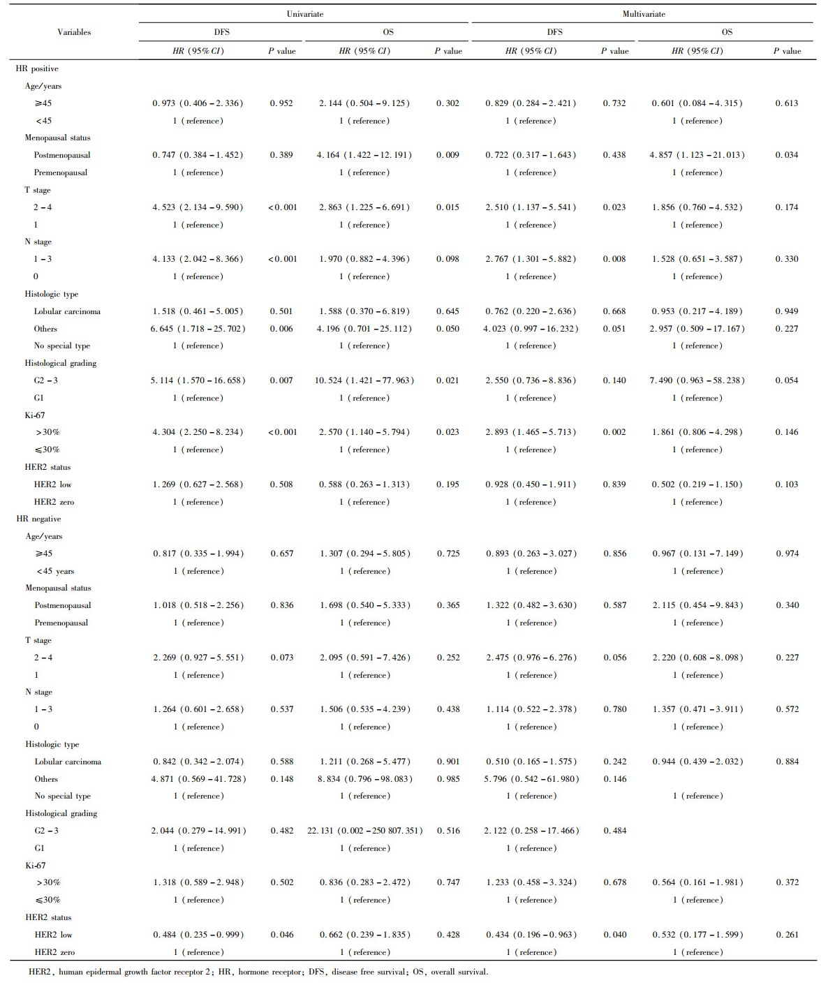
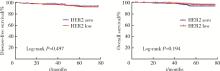
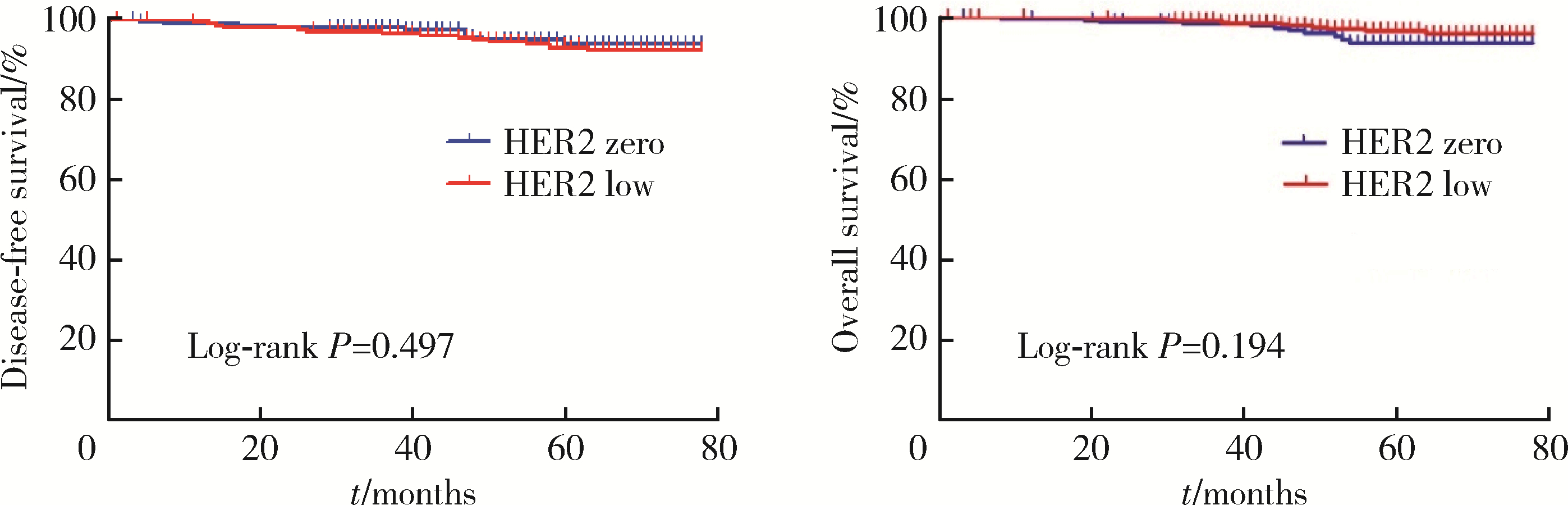
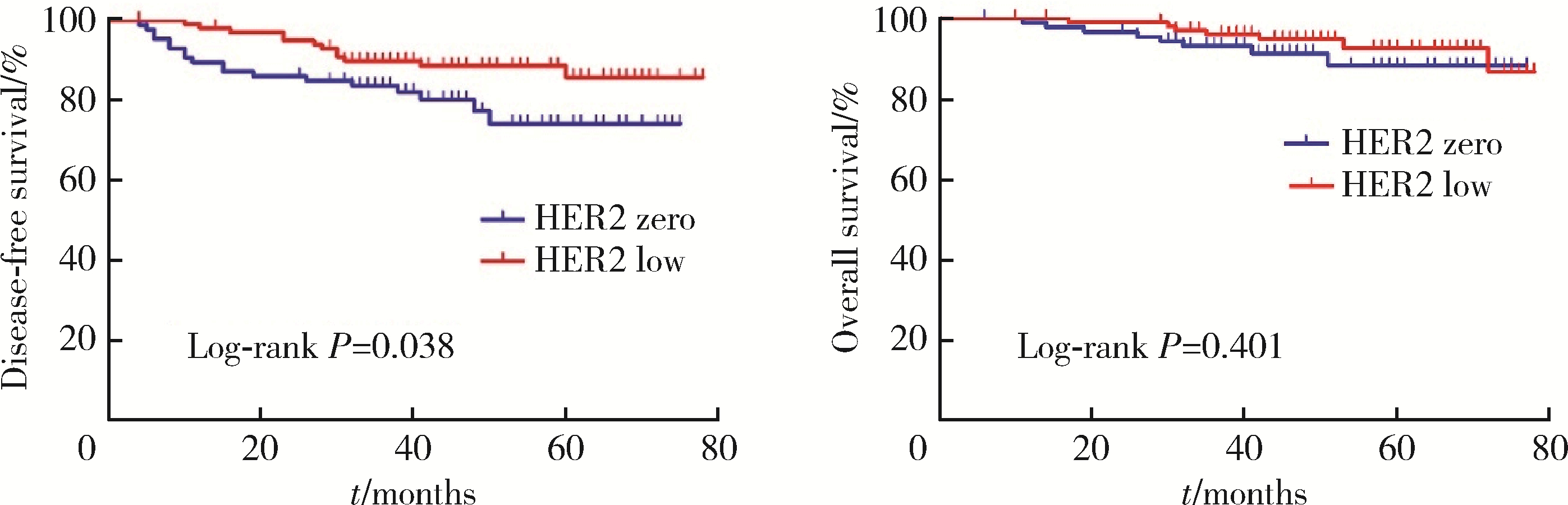
目的: 新型抗人表皮生长因子受体2(human epidermal growth factor receptor 2, HER2)抗体偶联药物临床试验的成功, 使HER2低表达乳腺癌是否将成为新的乳腺癌类型而受到关注。本研究分析比较HER2低表达与HER2零表达两组乳腺癌患者间的临床病理特征及生存数据, 探讨其临床生物学上的差异性。方法: 2014年1月至2017年12月北京大学第一医院乳腺疾病中心收治的1 250例女性原发性非转移性乳腺癌中, 969例HER2阴性(免疫组织化学染色评分为0、1+、2+, 且荧光原位杂交未扩增), 分析其中HER2低表达(1+或2+且荧光原位杂交未扩增)和HER2零表达(0分)两组患者间临床病理特征及预后情况。对两组患者进行无病生存期(disease free survival, DFS)和总生存期(overall survival, OS)预后评价, 采用Kaplan-Meier曲线计算生存率, Log-rank检验比较生存差异, 单因素和多因素Cox回归分析预后影响因素。采用双侧检验, P < 0.05为差异有统计学意义。结果: 969例HER2阴性乳腺癌中, HER2低表达者606例(62.54%)、HER2零表达者363例(37.46%)。与HER2零表达组相比, HER2低表达组的N分期(P=0.001)和TNM分期更高(P=0.044), 非特殊型组织学类型占比(82.7% vs. 79.1%, P=0.009)和组织学分级更高(P=0.048), 激素受体阳性率偏高(83.2% vs. 75.2%, P=0.003), Ki-67增殖指数>30%者的比例偏低(30.4% vs. 36.6%, P=0.044)。两组间DFS及OS差异无统计学意义(P>0.05)。969例患者中, 激素受体阳性777例, 激素受体阴性(即三阴性乳腺癌)192例。激素受体阳性的777例中, HER2低表达504例(64.9%), HER2零表达273例(35.1%), 两组比较, HER2低表达组的发病年龄小(P=0.016), 未绝经者占比高(P=0.029), 淋巴结受累更多(P=0.002), TNM分期更高(P=0.031), 小叶癌和黏液癌组织学类型较少见(3.6% vs. 7.3%, 4.8% vs. 10.6%, P < 0.001), DFS和OS差异无统计学意义(P>0.05)。192例激素受体阴性(三阴性乳腺癌)中, HER2低表达102例(53.1%), HER2零表达90例(46.9%), 两组比较, HER2低表达组的发病年龄大(P=0.001), 未绝经所占比例低(P=0.029), 组织学分级更低(P < 0.001), Ki-67增殖指数更低(P < 0.001), 雄激素受体阳性率高(58.8% vs. 34.4%, P < 0.001), DFS比较好(P=0.038), OS差异无统计学意义(P>0.05)。结论: HER2低表达乳腺癌占所有乳腺癌约一半的比例, 发病远高于HER2阳性乳腺癌, 其临床病理特征存在异质性, 激素受体表达状态对HER2低表达乳腺癌的临床生物学有影响。

关键词: 乳腺癌, 人表皮生长因子受体2, 低表达, 临床病理特征, 预后

Abstract:

Objective: There is an increasing interest in human epidermal growth factor receptor 2 (HER2) low expression breast cancer with the result of novel anti-HER2 antibody-drug conjugates for breast cancer. HER2 low expression breast cancer is expected to become a new type of breast cancer. This study analyzed and compared the clinicopathological features and survival data of breast cancer with HER2 low expression group [immunohistochemistry (IHC) 1+ or IHC 2+, and fluorescence in situ hybridization (FISH) negative] and HER2 zero expression group (IHC 0), in order to explore the difference in clinical biology of HER2 low expression breast cancers. Methods: Among 1 250 female patients with primary non-metastatic breast cancer admitted to the Breast Disease Center of Peking University First Hospital from January 2014 to December 2017, 969 cases were HER2 negative (IHC 0, 1+, 2+, and FISH was not amplified). The clinicopathologic features and prognosis of the patients with HER2 low expression (IHC 1+ or 2+, and unamplified by FISH) and HER2 zero expression (IHC 0) were analyzed. Disease free survival (DFS) and overall survival (OS) were evaluated, survival rates were calculated by Kaplan-Meier curve, and survival differences were compared by Log-rank test. Cox regression analysis of univariate and multivariate prognostic factors. Bilateral test was used, and P < 0.05 was considered statistically significant. Results: In the 969 patients with HER2 negative breast cancer, 606 had HER2 low expression (62.54%) and 363 had HER2 zero expression (37.46%). Compared with breast cancer with HER2 zero expression, those with HER2 low expression had higher N stage (P=0.001) and TNM stage (P=0.044), the proportion of non-specific histological types was higher (82.7% vs. 79.1%, P=0.009), the histological grade was higher (P=0.048), and the positive rate of hormone receptor was higher (83.2% vs. 75.2%, P=0.003). The percentage of Ki-67 value index >30% was lower (30.4% vs. 36.6%, P=0.044). There was no significant difference in DFS and OS between the two groups (P>0.05). In the 969 cases, 777 were hormone receptor positive and 192 were hormone receptor negative (triple negative cancer). Among the 777 cases with hormone receptor positive, 504 (64.9%) were HER2 low expression, and 273 (35.1%) were HER2 zero expression. Compared with breast cancer with HER2 zero expression group, the HER2 low expression group had a younger age (P=0.016), a higher proportion of premenopausal patients (P=0.029), more lymph node involvement (P=0.002), and a higher total TNM stage (P=0.031), and less frequent histological types of lobular and mucinous carcinoma (3.6% vs. 7.3%, 4.8% vs. 10.6%, P=0.001). There was no difference in DFS and OS between HER2 low expression and zero expression (P>0.05). Among 192 patients with hormone receptor negative, there were 102 cases (53.1%) with HER2 low expression and 90 cases (46.9%) with HER2 zero expression. Compared with the HER2 zero expression groups, HER2 low expression group was older (P=0.001), the proportion of premenopausal patients was low (P=0.029), the histological grade was lower (P < 0.001), the Ki-67 value index was lower (P < 0.001), and androgen receptor positive rate was higher (58.8% vs. 34.4%, P < 0.001). DFS was better than HER2 zero expression group (P=0.038), but there was no difference in OS between the two groups (P>0.05). Conclusion: HER2 low expression breast cancer accounts for about half of all breast cancers, and the incidence is much higher than that of HER2 positive breast cancer. Its clinicopathologic features are heterogeneous, and the status of hormone receptor expression has an impact on the clinical biology of this group.

Key words: Breast cancer, Human epidermal growth factor receptor 2, Low expression, Clinicopathological features, Prognosis

中图分类号: 

  • R737.9

表1

HER2低表达与HER2零表达乳腺癌患者的临床特征分析"

Characteristics All cases (n=969) HR positive cases (n=777) HR negative cases (n=192)
HER2 zero HER2 low P value HER2 zero HER2 low P value HER2 zero HER2 low P value
Age/years
    Total 55 (46, 65) 55 (46, 64) 0.545 57 (48, 66) 54 (45, 64) 0.016 52 (42, 62) 58 (50, 67) 0.001
    < 45 64 (17.6) 104 (17.2) 38 (13.9) 96 (19.0) 26 (28.9) 8 (7.8)
    ≥45 299 (82.4) 502 (82.8) 235 (86.1) 408 (81.0) 64 (71.1) 94 (92.2)
Menopausal status
    Postmenopausal 213 (58.7) 342 (56.4) 0.495 105 (38.5) 235 (46.6) 0.029 45 (50.0) 45 (50.0) 0.029
    Premenopausal 150 (41.3) 264 (43.6) 168 (61.5) 269 (53.4) 73 (71.6) 29 (28.4)
T stage
    1 199 (54.8) 314 (51.8) 0.758 166 (60.8) 279 (55.4) 0.145 33 (36.7) 35 (34.3) 0.943
    2 150 (41.3) 273 (45.0) 100 (36.6) 210 (41.7) 50 (55.6) 63 (61.8)
    3 12 (3.3) 13 (2.1) 6 (2.2) 11 (2.2) 6 (6.7) 2 (2.0)
    4 2 (0.6) 6 (1.0) 1 (0.4) 4 (0.8) 1 (1.1) 2 (2.0)
N stage
    0 255 (70.2) 360 (59.4) 0.001 192 (70.3) 293 (59.1) 0.002 63 (70.0) 67 (65.7) 0.341
    1 80 (22.0) 182 (30.0) 59 (21.6) 162 (32.1) 21 (23.3) 20 (19.6)
    2 19 (5.2) 44 (7.3) 15 (5.5) 36 (7.1) 4 (4.4) 8 (7.8)
    3 9 (2.5) 20 (3.3) 7 (2.6) 13 (2.6) 2 (2.2) 7 (6.9)
TNM stage
    Ⅰ 163 (44.9) 237 (39.1) 0.044 137 (50.2) 210 (41.7) 0.031 26 (28.9) 27 (26.5) 0.207
    Ⅱ 167 (46.0) 295 (48.7) 110 (40.3) 238 (47.2) 57 (63.3) 57 (55.9)
    Ⅲ 33 (9.1) 74 (12.2) 26 (9.5) 56 (11.1) 7 (7.8) 18 (17.6)

表2

HER2低表达与HER2零表达乳腺癌患者的病理特征分析"

Characteristics All cases (n=969) HR positive cases (n=777) HR negative cases (n=192)
HER2 zero HER2 low P value HER2 zero HER2 low P value HER2 zero HER2 low P value
Histologic type, n (%)
    No special type 287 (79.1) 501 (82.7) 0.009 212 (77.7) 423 (83.9) < 0.001 75 (83.3) 77 (75.5) 0.306
    Lobular carcinoma 20 (5.5) 20 (3.3) 20 (7.3) 18 (3.6) 0 (0) 2 (2.0)
    Mucinous carcinoma 29 (8.0) 24 (4.0) 29 (10.6) 24 (4.8) 0 (0) 0 (0)
    Others 27 (7.4) 61 (10.1) 12 (4.4) 39 (7.7) 15 (16.7) 22 (21.6)
Histological grading, n (%)
    G1 82 (22.8) 156 (25.8) 0.048 73 (29.3) 146 (29.0) 0.448 3 (3.3) 10 (9.9) < 0.001
    G2 166 (46.1) 299 (49.4) 149 (55.2) 260 (51.6) 17 (18.9) 39 (38.6)
    G3 112 (31.1) 150 (24.8) 42 (15.6) 98 (19.4) 70 (77.8) 52 (51.5)
    Absent 3 (0.8) 1 (0.2)
Ki-67, n (%)
    ≤30% 230 (63.4) 422 (69.6) 0.044 214 (78.4) 377 (74.8) 0.263 16 (17.8) 45 (44.1) < 0.001
   >30% 133 (36.6) 184 (30.4) 59 (21.6) 127 (25.2) 74 (82.2) 57 (55.9)

表3

HER2阴性乳腺癌中HER2状态与激素受体表达之间的关系"

Items HER2 zero, n (%) HER2 low, n (%) P value
HR positive 273 (75.2) 504 (83.2) 0.003
HR negative 90 (24.8) 102 (16.8)

表4

激素受体阴性乳腺癌中HER2状态与CK5/6和/或EGFR、AR表达之间的关系"

Items HER2 zero, n (%) HER2 low, n (%) P value
CK5/6 and/or EGFR
    CK5/6 and/or EGFR positive 77 (85.6) 74 (72.5)
    CK5/6 and/or EGFR negative 10 (11.1) 20 (24.5) 0.056
    Absent 3 (3.3) 3 (2.9)
AR
    AR positive 31 (34.4) 60 (58.8)
    AR negative 51 (56.7) 27 (26.5) < 0.001
    Absent 8 (8.9) 15 (14.7)

表5

HER2阴性乳腺癌患者的预后因素分析"

图1

HER2零表达和HER2低表达乳腺癌患者的DFS和OS生存曲线比较"

表6

激素受体不同状态HER2阴性乳腺癌患者的预后因素分析"

图2

HER2零表达和HER2低表达激素受体阳性乳腺癌患者的DFS和OS生存曲线比较"

图3

HER2零表达和HER2低表达激素受体阴性乳腺癌患者的DFS和OS生存曲线比较"

1 Yarden Y . Biology of HER2 and its importance in breast cancer[J]. Oncology, 2001, 61 (Suppl 2): 1- 13.
2 Choong GM , Cullen GD , O'Sullivan CC , et al. Evolving stan-dards of care and new challenges in the management of HER2-positive breast cancer[J]. CA Cancer J Clin, 2020, 70 (5): 355- 374.
doi: 10.3322/caac.21634
3 Swain SM , Baselga J , Kim SB , et al. Pertuzumab, trastuzumab, and docetaxel in HER2-positive metastatic breast cancer[J]. N Engl J Med, 2015, 372 (8): 724- 734.
doi: 10.1056/NEJMoa1413513
4 Wolff AC , Hammond ME , Hicks DG , et al. Recommendations for human epidermal growth factor receptor 2 testing in breast cancer: American Society of Clinical Oncology/College of American Patho-logists clinical practice guideline update[J]. J Clin Oncol, 2013, 31 (31): 3997- 4013.
doi: 10.1200/JCO.2013.50.9984
5 Gilcrease MZ , Woodward WA , Nicolas MM , et al. Even low-level HER2 expression may be associated with worse outcome in node-positive breast cancer[J]. Am J Surg Pathol, 2009, 33 (5): 759- 767.
doi: 10.1097/PAS.0b013e31819437f9
6 Modi S , Park H , Murthy RK , et al. Antitumor activity and safety of trastuzumab deruxtecan in patients with HER2-low-expressing advanced breast cancer: Results from a phase Ⅰb study[J]. J Clin Oncol, 2020, 38 (17): 1887- 1896.
doi: 10.1200/JCO.19.02318
7 Tarantino P , Hamilton E , Tolaney SM , et al. HER2-low breast cancer: Pathological and clinical landscape[J]. J Clin Oncol, 2020, 38 (17): 1951- 1962.
doi: 10.1200/JCO.19.02488
8 Hammond ME , Hayes DF , Dowsett M , et al. American Society of Clinical Oncology/College Of American Pathologists guideline recommendations for immunohistochemical testing of estrogen and progesterone receptors in breast cancer[J]. J Clin Oncol, 2010, 28 (16): 2784- 2795.
doi: 10.1200/JCO.2009.25.6529
9 Schalper KA , Kumar S , Hui P , et al. A retrospective population-based comparison of HER2 immunohistochemistry and fluorescence in situ hybridization in breast carcinomas: Impact of 2007 American Society of Clinical Oncology/College of American Pathologists criteria[J]. Arch Pathol Lab Med, 2014, 138 (2): 213- 219.
doi: 10.5858/arpa.2012-0617-OA
10 Xin L , Wu Q , Zhan C , et al. Multicenter study of the clinico-pathological features and recurrence risk prediction model of early-stage breast cancer with low-positive human epidermal growth factor receptor 2 expression in China (Chinese Society of Breast Surgery 021)[J]. Chin Med J (Engl), 2022, 135 (6): 697- 706.
doi: 10.1097/CM9.0000000000002056
11 Schettini F , Chic N , Brasó-Maristany F , et al. Clinical, patholo-gical, and PAM50 gene expression features of HER2-low breast cancer[J]. NPJ Breast Cancer, 2021, 7 (1): 1.
doi: 10.1038/s41523-020-00208-2
12 Denkert C , Seither F , Schneeweiss A , et al. Clinical and molecular characteristics of HER2-low-positive breast cancer: Pooled analysis of individual patient data from four prospective, neoadjuvant clinical trials[J]. Lancet Oncol, 2021, 22 (8): 1151- 1161.
doi: 10.1016/S1470-2045(21)00301-6
13 Rossi V , Sarotto I , Maggiorotto F , et al. Moderate immunohistochemical expression of HER-2 (2+) without HER-2 gene amplification is a negative prognostic factor in early breast cancer[J]. Oncologist, 2012, 17 (11): 1418- 1425.
doi: 10.1634/theoncologist.2012-0194
14 Won HS , Ahn J , Kim Y , et al. Clinical significance of HER2-low expression in early breast cancer: A nationwide study from the Korean Breast Cancer Society[J]. Breast Cancer Res, 2022, 24 (1): 22.
doi: 10.1186/s13058-022-01519-x
15 Horisawa N , Adachi Y , Takatsuka D , et al. The frequency of low HER2 expression in breast cancer and a comparison of prognosis between patients with HER2-low and HER2-negative breast cancer by HR status[J]. Breast Cancer, 2022, 29 (2): 234- 241.
doi: 10.1007/s12282-021-01303-3
16 Dehghani M , Keshavarz P , Talei A , et al. The Effects of Low HER2/neu expression on the clinicopathological characteristics of triple-negative breast cancer patients[J]. Asian Pac J Cancer Prev, 2020, 21 (10): 3027- 3032.
doi: 10.31557/APJCP.2020.21.10.3027
17 Jacot W , Maran-Gonzalez A , Massol O , et al. Prognostic value of HER2-low expression in non-metastatic triple-negative breast cancer and correlation with other biomarkers[J]. Cancers (Basel), 2021, 13 (23): 6059.
doi: 10.3390/cancers13236059
[1] 赖玉梅,李忠武,李欢,吴艳,时云飞,周立新,楼雨彤,崔传亮. 68例肛管直肠黏膜黑色素瘤临床病理特征及预后[J]. 北京大学学报(医学版), 2023, 55(2): 262-269.
[2] 沈棋,刘亿骁,何群. 肾黏液样小管状和梭形细胞癌的临床病理特点及预后[J]. 北京大学学报(医学版), 2023, 55(2): 276-282.
[3] 宿骞,彭歆,周传香,俞光岩. 369例口腔颌面部非霍奇金淋巴瘤的临床病理特点及预后[J]. 北京大学学报(医学版), 2023, 55(1): 13-21.
[4] 张铨,宋海峰,马冰磊,张喆楠,周朝晖,李傲林,刘军,梁磊,朱时雨,张骞. 术前预后营养指数可作为预测非转移性肾细胞癌预后的指标[J]. 北京大学学报(医学版), 2023, 55(1): 149-155.
[5] 邢晓燕,张筠肖,朱冯赟智,王一帆,周新尧,李玉慧. 皮肌炎合并巨噬细胞活化综合征5例[J]. 北京大学学报(医学版), 2022, 54(6): 1214-1218.
[6] 曹乐清,周婧睿,陈育红,陈欢,韩伟,陈瑶,张圆圆,闫晨华,程翼飞,莫晓冬,付海霞,韩婷婷,吕萌,孔军,孙于谦,王昱,许兰平,张晓辉,黄晓军. 异基因造血干细胞移植后晚发重症肺炎患者治疗与预后转归的关系[J]. 北京大学学报(医学版), 2022, 54(5): 1013-1020.
[7] 方伟岗,田新霞,解云涛. 基因多态性对中国汉族女性乳腺癌遗传易感性的影响[J]. 北京大学学报(医学版), 2022, 54(5): 822-828.
[8] 王跃,张爽,张虹,梁丽,徐玲,程元甲,段学宁,刘荫华,李挺. 激素受体阳性/人表皮生长因子受体2阴性乳腺癌临床病理特征及预后[J]. 北京大学学报(医学版), 2022, 54(5): 853-862.
[9] 张崔建,何志嵩,周利群. 上尿路尿路上皮癌的淋巴清扫[J]. 北京大学学报(医学版), 2022, 54(4): 592-594.
[10] 邱敏,连岩岩,陆敏,王滨帅,田晓军,卢剑,刘承,张树栋,姜敏,马潞林. 多原发癌合并肾癌的治疗及预后[J]. 北京大学学报(医学版), 2022, 54(4): 680-685.
[11] 博尔术,洪鹏,张宇,邓绍晖,葛力源,陆敏,李楠,马潞林,张树栋. 乳头状肾细胞癌的临床病理特征和预后分析[J]. 北京大学学报(医学版), 2022, 54(4): 615-620.
[12] 邓宇含,姜勇,王子尧,刘爽,汪雨欣,刘宝花. 基于长短期记忆网络和Logistic回归的重症监护病房脑卒中患者院内死亡风险预测[J]. 北京大学学报(医学版), 2022, 54(3): 458-467.
[13] 丁婷婷,曾楚雄,胡丽娜,余明华. 基于癌症基因组图谱数据库结直肠癌免疫细胞浸润预测模型的建立[J]. 北京大学学报(医学版), 2022, 54(2): 203-208.
[14] 蓝璘,贺洋,安金刚,张益. 颧骨缺损不同修复重建方法和预后的回顾性分析[J]. 北京大学学报(医学版), 2022, 54(2): 356-362.
[15] 王飞,朱翔,贺蓓,朱红,沈宁. 自发缓解的滤泡性细支气管炎伴非特异性间质性肺炎1例报道并文献复习[J]. 北京大学学报(医学版), 2021, 53(6): 1196-1200.
Viewed
Full text


Abstract

Cited

  Shared   
  Discussed   
[1] 王军, 肖水芳, 秦永, 王全桂, 陈丽. 以面神经麻痹为首诊表现的Wegener肉芽肿病一例[J]. 北京大学学报(医学版), 2007, 39(4): 434 -436 .
[2] 柳晓辉, 那加, 刘玲玲, 罗斌. 头颈部血管肉瘤3例[J]. 北京大学学报(医学版), 2001, 33(3): 288 -289 .
[3] 张震康. 口腔医学科学研究的重要进展和方向[J]. 北京大学学报(医学版), 2002, 34(2): 97 -98 .
[4] 梁成, 王兴, 伊彪, 李自力, 王晓霞. 骨性颞下颌关节强直伴小颌畸形及阻塞性睡眠呼吸暂停综合征的牵引成骨治疗[J]. 北京大学学报(医学版), 2002, 34(2): 112 -116 .
[5] 张勇, 栾庆先. 牙周维护治疗在保持牙周长期疗效中的作用[J]. 北京大学学报(医学版), 2011, 43(1): 29 -33 .
[6] 夏永华, 刘冬, 张彩凤, 付丹丹, 李敏, 李占国, 田中伟. NF-κB信号通路的阻断对皮肤鳞癌SCL-1细胞凋亡的影响[J]. 北京大学学报(医学版), 2011, 43(2): 179 -182 .
[7] 孙宇, 刘毅强, 冯国双, 李吉友. 转化生长因子β1在萎缩性胃炎发生中的作用[J]. 北京大学学报(医学版), 2009, 41(6): 635 -639 .
[8] 张少衡, 贾竹青, 郭静萱, 张萍, 马康涛, 王淑玲, 刘永刚, 李凌松, 周春燕. 骨髓细胞移植上调血管内皮生长因子及其受体的表达并改善缺血心脏功能[J]. 北京大学学报(医学版), 2003, 35(4): 429 -433 .
[9] 李文海, 张建中. 二期梅毒皮疹中梅毒螺旋体基因检测和浸润细胞研究[J]. 北京大学学报(医学版), 2003, 35(5): 485 -487 .
[10] 李海霞, 屈晨雪, 徐国宾, 闫存玲, 张国华, 李传保, 王建中, 夏铁安. 检验科SARS标本检测安全管理介绍[J]. 北京大学学报(医学版), 2003, 35(z1): 92 -94 .