北京大学学报(医学版) ›› 2018, Vol. 50 ›› Issue (6): 1083-1087. doi: 10.19723/j.issn.1671-167X.2018.06.025
茶溶液着色离体牛切牙的着色效果
朱曚曚1,李应龙2,潘洁1,△( )
)
- 1. 北京大学口腔医学院?口腔医院, 综合科 国家口腔疾病临床医学研究中心 口腔数字化医疗技术和材料国家工程实验室 口腔数字医学北京市重点实验室, 北京 100081
2. 北京大学前沿交叉学科研究院, 北京 100871
Staining effect of bovine incisors in vitro by tea solutions
Meng-meng ZHU1,Ying-long LI2,Jie PAN1,△()
- 1. Department of General, Peking University School and Hospital of Stomatology & National Clinical Research Center for Oral Diseases & National Engineering Laboratory for Digital and Material Technology of Stomatology & Beijing Key Laboratory of Digital Stomatology, Beijing 100081, China
2. Academy for Advanced Interdisciplinary Studies, Peking University, Beijing 100871, China
摘要:
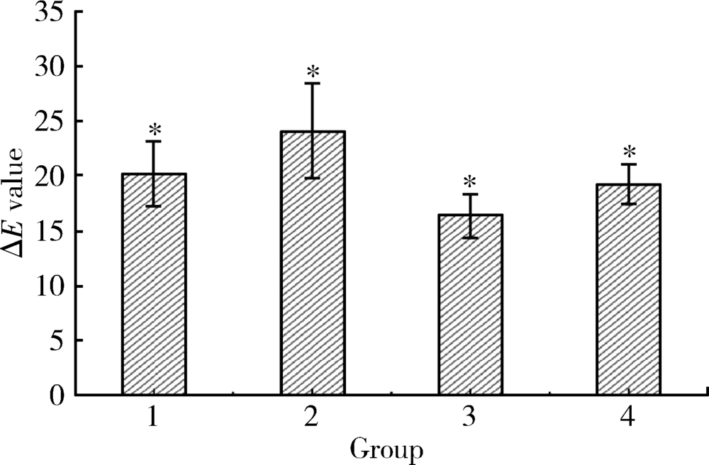
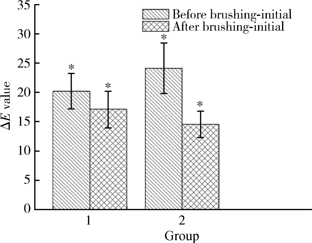
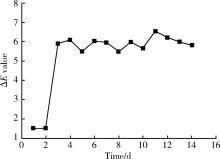
目的: 比较不同浓度、不同着色方式的茶溶液着色离体牛切牙的效果及着色的持久性,探究茶溶液着色离体牙模型的最佳条件。方法: 选用牙冠唇面中1/3比色为A1色的牛切牙20颗,纵剖为40个样本,随机分为4组(n=10)。1组:2%(质量分数)茶溶液连续着色6 d;2组:2%茶溶液着色6 d,每日换液;3组:1%茶溶液连续着色6 d;4组:1%茶溶液着色6 d,每日换液。6 d后,经Crystaleye比色仪测量牙冠唇面中1/3的Lab值并计算ΔE值,选出着色效果最佳的浓度组,使用国际通用标准牙刷切龈向轻刷牙面30次,测量并计算ΔE值,选出着色效果最佳的换液方式组观察其稳定性,将样本浸泡于人工唾液中37 ℃保存14 d,每天测量并计算ΔE值,选出着色效果稳定的时间。结果: 着色6 d后,换液方式相同时,2%茶溶液着色效果优于1%组(20.21 vs. 16.44, 24.09 vs. 19.22, P<0.05);茶溶液浓度相同时,每天换液组优于不换液组(24.09 vs. 20.21, 19.22 vs. 16.44, P<0.05)。选1、2组进行刷牙实验,刷牙后两组颜色均变浅,且不换液组着色效果优于每天换液组(3.06 vs. 9.51, P<0.05)。选择着色效果最好的2%茶溶液连续浸泡6天组,放入人工唾液浸泡14 d,第1、2天牙面颜色无明显变化(1.51 vs. 1.51, P>0.05),第3天明显变浅(1.51 vs. 5.89, P<0.05), 至第14天颜色再无明显变化(5.89 vs. 5.81, P>0.05),且深于着色前。结论: 将离体牙在2%茶溶液连续着色6 d,然后将其保存在人工唾液中,第3~14天内着色效果稳定,可作为理想的离体牙着色模型。
中图分类号:
- R781



| [1] |
Rajesh AG, Ranganath LM, Kumar KS , et al. Surface morpho-logical changes in human enamel following bleaching: an in vitro scanning electron microscopic study[J]. J Contemp Dent Pract, 2012,13(3):405-415.
doi: 10.5005/jp-journals-10024-1159 pmid: 22918018 |
| [2] |
Xing W, Jiang T, Liang S , et al. Effect of in-office bleaching agents on the color changes of stained ceromers and direct com-posite resins[J]. Acta Odontol Scand, 2014,72(8):1032-1038.
doi: 10.3109/00016357.2014.946962 pmid: 25196263 |
| [3] |
Lee BS, Huang SH, Chiang YC , et al. Development of in vitro tooth staining model and usage of catalysts to elevate the effectiveness of tooth bleaching[J]. Dent Mater, 2008,24(1):57-66.
doi: 10.1016/j.dental.2007.01.012 pmid: 17442388 |
| [4] |
Lee RJ, Bayne A, Tiangco M , et al. Prevention of tea-induced extrinsic tooth stain[J]. Int J Dent Hyg, 2014,12(4):267-272.
doi: 10.1111/idh.12096 pmid: 25040739 |
| [5] |
Agnihotry A, Gill KS, Singhal D , et al. A Comparison of the bleaching effectiveness of chlorine dioxide and hydrogen peroxide on dental composite[J]. Braz Dent J, 2014,25(6):524-527.
doi: 10.1590/0103-6440201300098 pmid: 25590199 |
| [6] |
Homayoom A, Sadat HKS, Nafiseh Z , et al. In vitro effect of casein phosphopeptide amorphous calcium phosphate on enamel susceptibility to staining by tea during bleaching treatment[J]. J Dent, 2015,12(8):607-613.
pmid: 27123021 |
| [7] |
Çelik C, Yüzügüllü B, Erkut S , et al. Effect of bleaching on staining susceptibility of resin composite restorative materials[J]. J Esthet Restor Dent, 2010,21(6):407-414.
doi: 10.1111/j.1708-8240.2009.00299.x pmid: 20002929 |
| [8] |
Suliemana M, Addya M, Macdonalda E , et al. The bleaching depth of a 35% hydrogen peroxide based in-office product: a study in vitro[J]. J Dent, 2005,33(1):33-40.
doi: 10.1016/j.jdent.2004.07.008 |
| [9] |
Sulieman M, Addy M, Rees JS . Development and evaluation of a method in vitro to study the effectiveness of tooth bleaching[J]. J Dent, 2003,31(6):415-422.
doi: 10.1016/S0300-5712(03)00069-1 pmid: 12878024 |
| [10] | 路丽 . 着色牙外漂白后再矿化的效果及对细菌粘附影响的体外实验研究[D]. 山东: 山东大学, 2005. |
| [11] | 李艳 . 三种着色牙模型漂白前后颜色的比较研究[D]. 湖南: 中南大学, 2012. |
| [12] |
Odaira C, Itoh S, Ishibashi K . Clinical evaluation of a dental color analysis system: the crystaleyespectrophotometer[J]. J Prosthodont Res, 2011,55(4):199-205.
doi: 10.1016/j.jpor.2010.12.005 pmid: 21296639 |
| [13] |
Wang C, Li Y, Wang X , et al. The enamel microstructures of bovine Omandibular incisors[J]. Anat Rec, 2012,295(10):1698-1706.
doi: 10.1002/ar.22543 pmid: 22837182 |
| [14] |
Addy M, Moran J . Mechanism of stain formation on teeth, in particular associated with metal ions and antiseptics[J]. Adv Dent Res, 1995,9(4):450-456.
doi: 10.1177/08959374950090041601 |
| [15] |
Carpenter GH, Pramanik R, Proctor GB . An in vitro model of chlorhexidine-induced tooth staining[J]. J Periodental Res, 2005,40(3):225-230.
doi: 10.1111/j.1600-0765.2005.00791.x pmid: 15853968 |
| [16] |
Berger SB, Coelho AS, Oliveira VA , et al. Enamel susceptibility to red wine staining after 35% hydrogen peroxide bleaching[J]. J Appl Oral Sci, 2008,16(3):201-204.
doi: 10.1590/S1678-77572008000300007 |
| [17] |
Phan X, Akyalcin S, Wiltshire WA , et al. Effect of tooth bleaching on shear bond strength of a fluoride- releasing sealant[J]. Angle Orthod, 2012,82(3):546-551.
doi: 10.2319/052711-353.1 pmid: 21854189 |
| [18] |
Joiner A, Muller D, Elofsson UM , et al. Ellipsometry analysis of the in vitro adsorption of tea polyphenols onto salivary pellicles[J]. Eur J Oral Sci, 2004,112(6):510-515.
doi: 10.1111/j.1600-0722.2004.00166.x pmid: 15560834 |
| [19] |
Pontefract H, Courtney M, Smith S , et al. Development of methods to enhance extrinsic tooth discoloration for comparison of toothpastes I studies in vitro[J]. J Clin Periodontol, 2004,31(1):1-6.
doi: 10.1111/cpe.2004.31.issue-1 |
| [20] |
Li Q, Xu BT, Li R , et al. Quantitative evaluation of colour regression and mineral content change of bleached teeth[J]. J Dent, 2010,38(3):253-260.
doi: 10.1016/j.jdent.2009.11.005 pmid: 19945498 |
| [21] |
李青, 王贻宁, 徐东选 . 四种常见离体牙保存液对牙齿颜色的影响. 口腔医学研究, 2005,21(2):197-199.
doi: 10.3969/j.issn.1671-7651.2005.02.027 |
| [1] | 杨静, 许晓韵, 郑丹妮, 凌晓彤, 屈留洋, 柳登高. 544例慢性唾液腺炎的临床与影像学特点及病因分析[J]. 北京大学学报(医学版), 2026, 58(3): 650-657. |
| [2] | 王昕莹, 程雪原, 张孟钧, 李菲, 段晋瑜, 乔静. 浓缩生长因子联合引导性组织再生术治疗下颌磨牙根分叉病变的疗效[J]. 北京大学学报(医学版), 2026, 58(2): 372-379. |
| [3] | 丛馨, 苏家增, 吴立玲, 丁冲, 李巍, 俞光岩. 唾液腺非肿瘤性疾病诊治研究进展[J]. 北京大学学报(医学版), 2026, 58(1): 1-9. |
| [4] | 杨雨婷, 屈留洋, 郑丹妮, 凌晓彤, 许晓韵, 柳登高. 1 812例唾液腺结石患者的人口学特征和临床特点[J]. 北京大学学报(医学版), 2026, 58(1): 153-159. |
| [5] | 臧海玲, 梁宇红. 上颌第二磨牙慢性根尖周炎合并器械分离的根管再治疗1例[J]. 北京大学学报(医学版), 2026, 58(1): 214-219. |
| [6] | 潘莲菲, 李文静, 王瑞洋, 焦剑, 曹战强, 高丽, 释栋. 口服抗生素辅助牙周机械治疗对重度牙周炎的短期疗效及影响因素[J]. 北京大学学报(医学版), 2026, 58(1): 30-36. |
| [7] | 孙菲, 王翠, 李思琪, 危伊萍, 余日月, 胡文杰. 赤藓糖醇喷砂与超声治疗对种植体周黏膜炎疗效的随机对照临床研究[J]. 北京大学学报(医学版), 2026, 58(1): 37-42. |
| [8] | 唐仁韬, 杨流畅, 聂杰, 王晓燕. 无窦型与有窦型根管治疗后慢性根尖周炎根管外菌群的组成及差异[J]. 北京大学学报(医学版), 2026, 58(1): 43-49. |
| [9] | 何梓玉, 张辉, 陈智滨, 邢海霞, 潘洁. 1例成年猛性龋患者龈上菌斑中小韦荣球菌的分离及代谢特性[J]. 北京大学学报(医学版), 2026, 58(1): 50-59. |
| [10] | 池彦廷, 蒋鸿杰, 陈艳, 徐志秀, 李斌斌. 直接免疫荧光在口腔黏膜寻常型天疱疮诊断中的价值: 基于多指标联合分析的回顾性研究[J]. 北京大学学报(医学版), 2026, 58(1): 68-73. |
| [11] | 马保金, 李建华, 桑元华, 于洋, 仇吉川, 邵金龙, 李凯, 刘世岳, 杜密, 商玲玲, 葛少华. 基于微环境和干细胞调控的牙周组织再生关键技术的建立与应用[J]. 北京大学学报(医学版), 2025, 57(5): 841-846. |
| [12] | 曹沛, 栾庆先. 牙周炎与全身系统性疾病的思考与探索[J]. 北京大学学报(医学版), 2025, 57(5): 852-858. |
| [13] | 孙翔宇, 袁超, 周芯竹, 刁婧, 郑树国. 唾液微生态在口腔及全身疾病早期防治中的应用[J]. 北京大学学报(医学版), 2025, 57(5): 859-863. |
| [14] | 包振英, 王雅杰. 炎症指标和细胞因子联合检测在慢性牙周炎中的应用[J]. 北京大学学报(医学版), 2025, 57(4): 772-778. |
| [15] | 朱慧, 闵赛南, 苏家增, 陈艳, 彭歆, 于尧, 俞光岩. 口腔黏膜嗜酸性溃疡的临床病理分析[J]. 北京大学学报(医学版), 2025, 57(3): 620-625. |
|
||
