北京大学学报(医学版) ›› 2019, Vol. 51 ›› Issue (4): 753-757. doi: 10.19723/j.issn.1671-167X.2019.04.028
扫频光学相干断层扫描根管内窥影像系统的建立及其在根裂诊断的应用
戚苈源1,陈晨2,姜岚2,李嘉男3,△( ),梁宇红1,4,△()
- 1. 北京大学口腔医学院?口腔医院,牙体牙髓科 国家口腔疾病临床医学研究中心 口腔数字化医疗技术和材料国家工程实验室 口腔数字医学北京市重点实验室, 北京 100081
2. 北京大学口腔医学院?口腔医院第一门诊部, 北京 100034
3. 中国科学院西安光学精密机械研究所瞬态光学与光子技术国家重点实验室, 西安 710000
4. 北京大学国际医院口腔科, 北京 102206
Construction of swept source optical coherence tomography imaging system for root canal endoscopy and application in diagnosis of root fractures
Li-yuan QI1,Chen CHEN2,Lan JIANG2,Jia-nan LI3,△(),Yu-hong LIANG1,4,△()
- 1. Department of Cariology and Endodontology,Peking University School and Hospital of Stomatology & National Clinical Research Center for Oral Diseases & National Engineering Laboratory for Digital and Material Technology of Stomatology & Beijing Key Laboratory of Digital Stomatology,Beijing 100081,China
2. First Clinical Division,Peking University School and Hospital of Stomatology,Beijing 100034,China
3. State Key Laboratory of Transient Optics and Photonics,Xian Institute of Optics and Precision Mechanics,Chinese Academy of Sciences,Xian,Shanxi 710000,China
4. Department of Stomatology,Peking University International Hospital,Beijing 102206,China
摘要:
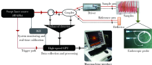
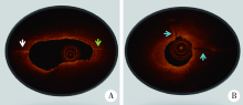
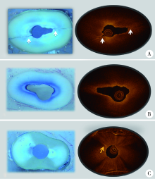
目的:建立扫频光学相干断层扫描(swept source-optical coherence tomography, SS-OCT)根管内窥影像系统,并评价该系统应用于诊断模拟根管内壁裂的准确性。方法:自主研发基于压电调谐滤波器并应用傅里叶(Fourier)域锁模技术构建的40 kHz超高速扫频激光光源系统(专利号200620135940.2),利用超微型梯度折射率透镜(专利号201320241218.7)制作极细根管内窥探头(直径0.86 mm),实现实时成像传输。构建的SS-OCT根管内窥影像系统扫频光源中心波长为1 310 nm,带宽为100 nm; 扫描图像的横向和纵向分辨率分别为25 μm和15 μm。利用人离体下颌前磨牙牙根制作人工模拟根裂(内壁裂),并制备高度1 mm的牙根横断面切盘。立体显微镜下观察发现,41个待测样本中有27个根管内壁裂样本(宽度在52~284 μm),另14个样本无根裂。应用上述构建的SS-OCT根管内窥影像系统扫描待测样本,重建图像的层厚为30 μm,层间距为30 μm。对1名口腔放射科医师和1名牙体牙髓科医师进行培训,判读SS-OCT扫描重建图像,判读根管内壁裂的有无及具体位置,评价两名观察者的自身一致性和观察者之间的一致性。以立体显微镜(组织学)检查结果作为金标准,评价应用SS-OCT根管内窥影像系统诊断模拟根管内壁裂的准确性。结果:两位观察者自身一致性的Kappa值分别为1.000和0.709,观察者之间的Kappa值为0.792。应用SS-OCT根管内窥影像系统扫描后27个根裂样本均被正确诊断,灵敏度为1.000,14个无根裂的样本有12个被正确判读,特异度为0.857, 2个无根裂样本被判读为有根裂,为假阳性。阳性预测值、阴性预测值分别为0.931、1.000,准确性为0.951。结论:扫频光学相干断层扫描根管内窥影像系统应用于观察根管内壁裂有临床应用前景。
中图分类号:
- R781



| [1] | Fercher AF, Drexler W, Hitzenberger CK , et al. Optical coherence tomography-principles and applications[J]. Rep Prog Phys, 2003,66(2):239. |
| [2] | Oliveira BPD , CÂmara AC, Duarte DA, et al. Detection of apical root cracks using spectral domain and swept-source optical co-herence tomography [J]. Int Endod J, 2017,43(7):1148-1151. |
| [3] | Lavinsky F, Lavinsky D . Novel perspectives on swept-source optical coherence tomography[J]. Int J Retina Vitreous, 2016,2(1):25. |
| [4] | Ha FJ, Giblett JP, Nerlekar N , et al. Optical coherence tomography guided percutaneous coronary intervention[J]. Heart Lung Circ, 2017,26(12):1267-1276. |
| [5] | Colston B, Everett M, Da Silva LB , et al. Imaging of hard-and soft-tissue structure in the oral cavity by optical coherence tomography[J]. Applied Optics, 1998,37(16):3582-3585. |
| [6] | Baumgartner A, Dichtl S, Hitzenberger CK , et al. Polarization-sensitive optical coherence tomography of dental structures[J]. Caries Res, 1999,34(1):59-69. |
| [7] | Shemesh H, van Soest G, Wu MK , et al. Diagnosis of vertical root fractures with optical coherence tomography[J]. J Endod, 2008,34(6):739-742. |
| [8] | Zain E, Zakian CM, Chew HP . Influence of the loci of non-cavitated fissure caries on its detection with optical coherence tomography[J]. J Dent, 2018,71(4):31-37. |
| [9] | Majkut P, Sadr A, Shimada Y , et al. Validation of optical coherence tomography against micro-computed tomography for evaluation of remaining coronal dentin thickness[J]. J Endod, 2015,41(8):1349-1352. |
| [10] | Han SH, Sadr A, Tagami J , et al. Non-destructive evaluation of an internal adaptation of resin composite restoration with swept-source optical coherence tomography and micro-CT[J]. Dent Mater, 2015,32(1):e1-e7 |
| [11] | Yoshioka T, Sakaue H, Ishimura H , et al. Detection of root surface fractures with swept-source optical coherence tomography (SS-OCT)[J]. Photomed Laser Surg, 2013,31(1):23-27. |
| [12] | 陈晨, 章文欣, 戚苈源 , 等. 光学相干断层扫描技术诊断牙根裂的实验研究[J]. 北京大学学报(医学版), 2018,50(3):547-552. |
| [13] | Wang P, Yan X, Liu D , et al. Detection of dental root fractures by using cone-beam computed tomography[J]. Dentomaxillofac Radiol, 2011,40(5):290-298. |
| [14] | Li G . Patient radiation dose and protection from cone-beam computed tomography[J]. Imaging Sci Dent, 2013,43(2):63-69. |
| [15] | Paul RA, Tamse A, Rosenberg E . Cracked and broken teeth definitions, differential diagnosis and treatment[J]. Refuat Hapeh Vehashinayim, 2007,24(2):7-12. |
| [16] | Huang D, Swanson EA, Lin CP , et al. Optical coherence tomography[J]. Science, 1991,254(5035):1178-1181 |
| [17] | Bahcall JK, Barss JT . Fiberoptic endoscope usage for intracanal visualization[J]. J Endod, 2001,27(2):128-129. |
| [18] | Hassan B, Metska ME, Ozok AR , et al. Detection of vertical root fractures in endodontically treated teeth by a cone beam computed tomography scan[J]. J Endod, 2009,35(5):719-722. |
| [19] | Özer SY . Detection of vertical root fractures of different thicknesses in endodontically enlarged teeth by cone beam computed tomography versus digital radiography[J]. J Endod, 2010,36(7):1245-1249. |
| [20] | Patel S, Brady E, Wilson R , et al. The detection of vertical root fractures in root filled teeth with periapical radiographs and CBCT scans[J]. Int Endod J, 2013,46(12):1140-1152. |
| [21] | Chavda R, Mannocci F, Andiappan M , et al. Comparing the in vivo diagnostic accuracy of digital periapical radiography with cone-beam computed tomography for the detection of vertical root fracture[J]. J Endod, 2014,40(10):1524-1529. |
| [22] | Makeeva IM, Byakova SF, Novozhilova NE , et al. Detection of artificially induced vertical root fractures of different widths by cone beam computed tomography in vitro and in vivo[J]. Int Endod J, 2016,49(10):980-989. |
| [1] | 杨静, 许晓韵, 郑丹妮, 凌晓彤, 屈留洋, 柳登高. 544例慢性唾液腺炎的临床与影像学特点及病因分析[J]. 北京大学学报(医学版), 2026, 58(3): 650-657. |
| [2] | 王昕莹, 程雪原, 张孟钧, 李菲, 段晋瑜, 乔静. 浓缩生长因子联合引导性组织再生术治疗下颌磨牙根分叉病变的疗效[J]. 北京大学学报(医学版), 2026, 58(2): 372-379. |
| [3] | 丛馨, 苏家增, 吴立玲, 丁冲, 李巍, 俞光岩. 唾液腺非肿瘤性疾病诊治研究进展[J]. 北京大学学报(医学版), 2026, 58(1): 1-9. |
| [4] | 杨雨婷, 屈留洋, 郑丹妮, 凌晓彤, 许晓韵, 柳登高. 1 812例唾液腺结石患者的人口学特征和临床特点[J]. 北京大学学报(医学版), 2026, 58(1): 153-159. |
| [5] | 臧海玲, 梁宇红. 上颌第二磨牙慢性根尖周炎合并器械分离的根管再治疗1例[J]. 北京大学学报(医学版), 2026, 58(1): 214-219. |
| [6] | 潘莲菲, 李文静, 王瑞洋, 焦剑, 曹战强, 高丽, 释栋. 口服抗生素辅助牙周机械治疗对重度牙周炎的短期疗效及影响因素[J]. 北京大学学报(医学版), 2026, 58(1): 30-36. |
| [7] | 孙菲, 王翠, 李思琪, 危伊萍, 余日月, 胡文杰. 赤藓糖醇喷砂与超声治疗对种植体周黏膜炎疗效的随机对照临床研究[J]. 北京大学学报(医学版), 2026, 58(1): 37-42. |
| [8] | 唐仁韬, 杨流畅, 聂杰, 王晓燕. 无窦型与有窦型根管治疗后慢性根尖周炎根管外菌群的组成及差异[J]. 北京大学学报(医学版), 2026, 58(1): 43-49. |
| [9] | 何梓玉, 张辉, 陈智滨, 邢海霞, 潘洁. 1例成年猛性龋患者龈上菌斑中小韦荣球菌的分离及代谢特性[J]. 北京大学学报(医学版), 2026, 58(1): 50-59. |
| [10] | 池彦廷, 蒋鸿杰, 陈艳, 徐志秀, 李斌斌. 直接免疫荧光在口腔黏膜寻常型天疱疮诊断中的价值: 基于多指标联合分析的回顾性研究[J]. 北京大学学报(医学版), 2026, 58(1): 68-73. |
| [11] | 马保金, 李建华, 桑元华, 于洋, 仇吉川, 邵金龙, 李凯, 刘世岳, 杜密, 商玲玲, 葛少华. 基于微环境和干细胞调控的牙周组织再生关键技术的建立与应用[J]. 北京大学学报(医学版), 2025, 57(5): 841-846. |
| [12] | 曹沛, 栾庆先. 牙周炎与全身系统性疾病的思考与探索[J]. 北京大学学报(医学版), 2025, 57(5): 852-858. |
| [13] | 孙翔宇, 袁超, 周芯竹, 刁婧, 郑树国. 唾液微生态在口腔及全身疾病早期防治中的应用[J]. 北京大学学报(医学版), 2025, 57(5): 859-863. |
| [14] | 包振英, 王雅杰. 炎症指标和细胞因子联合检测在慢性牙周炎中的应用[J]. 北京大学学报(医学版), 2025, 57(4): 772-778. |
| [15] | 朱慧, 闵赛南, 苏家增, 陈艳, 彭歆, 于尧, 俞光岩. 口腔黏膜嗜酸性溃疡的临床病理分析[J]. 北京大学学报(医学版), 2025, 57(3): 620-625. |
|
||
