北京大学学报(医学版) ›› 2020, Vol. 52 ›› Issue (1): 83-89. doi: 10.19723/j.issn.1671-167X.2020.01.013

• 论著 • 上一篇    下一篇

腮腺CT影像报告与数据系统的初步研究

李玉冰1,孙丽莎2,孙志鹏1,(),谢晓艳1,张建运3,张祖燕1,赵燕平1,马绪臣1   

  1. 1. 北京大学口腔医学院·口腔医院, 医学影像科, 北京 100081
    2. 北京大学口腔医学院·口腔医院,中心实验室, 北京 100081
    3. 北京大学口腔医学院·口腔医院,口腔病理科 国家口腔疾病临床医学研究中心 口腔数字化医疗技术和材料国家工程实验室 口腔数字医学北京市重点实验室, 北京 100081
  • 收稿日期:2019-10-10 出版日期:2020-02-18 发布日期:2020-02-20
  • 通讯作者: 孙志鹏 E-mail:sunzhipeng@bjmu.edu.cn

Parotid CT imaging reporting and data system: A preliminary study

Yu-bing LI1,Li-sha SUN2,Zhi-peng SUN1,(),Xiao-yan XIE1,Jian-yun ZHANG3,Zu-yan ZHANG1,Yan-ping ZHAO1,Xu-chen MA1   

  1. 1. Department of Oral Pathology, Peking University School and Hospital of Stomatology & Department of Oral and Maxillofacial Radiology, Beijing 100081, China
    2. Department of Oral Pathology, Peking University School and Hospital of Stomatology & Central Laboratory, Beijing 100081, China
    3. Department of Oral Pathology, Peking University School and Hospital of Stomatology & National Clinical Research Center for Oral Diseases & National Engineering Laboratory for Digital and Material Technology of Stomatology & Beijing Key Laboratory of Digital Stomatology, Beijing 100081, China
  • Received:2019-10-10 Online:2020-02-18 Published:2020-02-20
  • Contact: Zhi-peng SUN E-mail:sunzhipeng@bjmu.edu.cn

RICH HTML


摘要:

目的:初步建立腮腺CT影像报告与数据系统(Parotid Imaging Reporting and Data System,PI-RADS),并探讨其临床应用价值。方法:纳入2013年1月至2016年12月间因腮腺肿物就诊于北京大学口腔医院并进行手术治疗的病例,回顾性评估所有病例的影像资料,获取相关影像特征,评估肿瘤恶性风险概率,并分为6个等级(1级,正常腮腺; 2级,基本确定为良性病变或肿瘤; 3级,无明确恶性病变证据但不能确定为良性病变; 4级,怀疑为恶性肿瘤病变但证据不充分; 5级,恶性肿瘤影像征象较充分; 6级,有恶性肿瘤病理学证据)。结果:共纳入腮腺肿物病例897例次,其中良性病变905例次、恶性肿瘤98例次,影像诊断为2级、3级、4级和5级的病变中,恶性肿瘤的构成比分别为0.4%、5.7%、35.5%和96.7%,随PI-RADS分级呈逐渐增高趋势(Z=-15.579,P<0.001)。相邻等级[2级与3级(χ 2=12.048,P=0.001)、3级与4级(χ 2=75.231,P<0.001)、4级与5级(χ 2=32.266,P<0.001)]之间的恶性构成比差异有统计学意义。Cohen’s Kappa检验表明两位研究者分级诊断具有中度一致性(κ=0.614,P<0.001,95%CI: 0.569~0.695)。结论:应用影像诊断分级方法对腮腺肿瘤性疾病的诊断和临床治疗有一定的帮助。

关键词: 腮腺肿瘤, 体层摄影术, X线计算机, 诊断, 鉴别, 治疗

Abstract:

Objective: To establish a Parotid Imaging Reporting and Data System (PI-RADS) for CT diagnosis of the parotid gland neoplasms and to investigate the clinical applicable value and feasibility of PI-RADS. Methods: Patients who had been diagnosed with primary parotid gland neoplasms and had received surgical treatments in Peking University School and Hospital of Stomatology during the period of January 2013 to December 2016 were included in this study. The diagnoses were confirmed by the postoperative pathological examinations in all the patients. The CT imaging data of all patients were retrospectively reviewed and analyzed by two readers in consensus. Imaging characteristics related to the parotid neoplasms were extracted and quantified. Based on comprehensive analysis of the imaging characteristics, the probabilities of the benign and malignant neoplasms were evaluated and classified into six grades, PI-RADS 1-6 (PI-RADS 1: normal parotid gland; PI-RADS 2: confidently benign lesions; PI-RADS 3: probably benign lesions without confirmed evidence of malignancy; PI-RADS 4: suspected malignancy without sufficient evidence of malignancy; PI-RADS 5: confidently malignant lesions; PI-RADS 6: lesions with confirmed pathological evidence of malignancy). Results: A total of 897 patients with 1 003 parotid lesions were included. The lesions included 905 benign and 98 malignant lesions. The proportions of the malignancies in PI-RADS 2, PI-RADS 3, PI-RADS 4 and PI-RADS 5 according to the two readers in consensus were 0.4%, 5.7%, 35.5% and 96.7% respectively. The overall Cohen’s Kappa test showed medium consistency between the two independent researchers (κ=0.614, P<0.001, 95%CI: 0.569-0.695). Pearson Chi-square test showed that the proportions of malignancies increased with the diagnostic PI-RADS grades (Cochran-Armitage trend test, Z=-15.579, P<0.001). The results of Pearson Chi-square tests showed significant differences between the grades [PI-RADS 2 and 3 (χ 2=12.048, P=0.001); PI-RADS 3 and 4 (χ 2=75.231, P<0.001); PI-RADS 4 and 5 (χ 2=32.266, P<0.001)]. Conclusion: PI-RADS can be used to evaluate the risk of malignancy and will be helpful to improve the imaging diagnosis and clinical treatment of paro-tid gland neoplasms.

Key words: Parotid neoplasms, Tomography, X-ray computed, Diagnosis, differential, Therapy

中图分类号: 

  • R739.87

表1

腮腺肿物PI-RADS诊断结果与影像学征象"

Items PI-RADS 2 PI-RADS 3 PI-RADS 3b PI-RADS 4 PI-RADS 5 PI-RADS 6 Benign Malignancy
Size
Small (<2.0 cm) 30 82 7 7 2 0 118 10
Medium (2.0-3.0 cm) 158 453 4 49 14 1 624 55
Large (>3.0 cm) 56 102 1 20 14 3 163 33
Single/multiple
Single on one side 139 584 0 62 19 3 730 77
Multiple on one side 33 25 5 11 9 1 66 18
Multiple on two sides 72 28 7 3 2 0 109 3
Boundary
Absolutely clear 153 153 0 3 0 0 309 0
Majorly clear 68 261 2 21 4 0 345 11
Partially ill-defined 17 182 1 26 3 1 210 20
Majorly ill-defined 6 38 7 18 8 3 37 43
Ill-defined 0 3 2 8 15 0 4 24
Shape
Round 54 269 5 21 7 0 334 22
Oval 154 162 3 13 3 0 322 13
Scalloped 31 182 2 27 6 2 222 28
Irregular 5 24 2 15 14 2 27 35
Dominant density on plain CT
Fat 7 1 0 0 0 0 8 0
Liquid 30 59 0 4 12 0 91 2
Soft-tissue (partial fluid) 66 239 1 36 18 3 311 46
Soft-tissue 141 338 11 36 0 1 495 50
Enhancement degree on first enhanced phase
No significant 25 42 0 3 0 0 68 2
Slightly 10 122 0 12 4 0 140 8
Medium 29 203 5 32 13 1 236 47
Obvious 89 114 7 21 11 3 217 28
Significant 67 53 0 5 1 0 119 7
Enhancement pattern
Gradual 20 294 5 30 11 1 333 28
Fast in-out 124 128 1 23 7 2 254 31
Basically no enhancement 4 4 0 2 1 0 8 3
Gradual in-out 9 51 2 10 2 0 61 13
Enhancement distribution
Membrane enhancement 5 34 0 7 0 0 40 6
Even 152 182 12 24 15 1 346 36
Uneven to even 17 198 0 21 5 1 223 20
Uneven 14 84 0 17 8 2 99 26
No enhancement 32 36 0 4 0 0 72 0
Calcification
No 239 602 12 59 26 4 861 81
Small granular or capsule 5 31 0 10 1 0 38 9
Large irregular 0 4 0 7 3 0 6 8
Cystic percentage
0 177 488 12 49 26 2 683 71
≤25% 19 53 0 10 2 2 72 14
>25%, ≤50% 7 17 0 6 2 0 23 9
>50%, ≤75% 5 33 0 5 0 0 40 3
>75% 36 46 0 6 0 0 87 1
Adjacent structure destruction
No 237 609 7 64 16 3 867 69
Move 5 19 0 5 0 0 26 3
Poor boundary 2 8 5 6 9 0 12 18
Significant 0 1 0 1 5 1 0 8

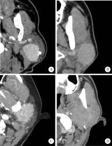
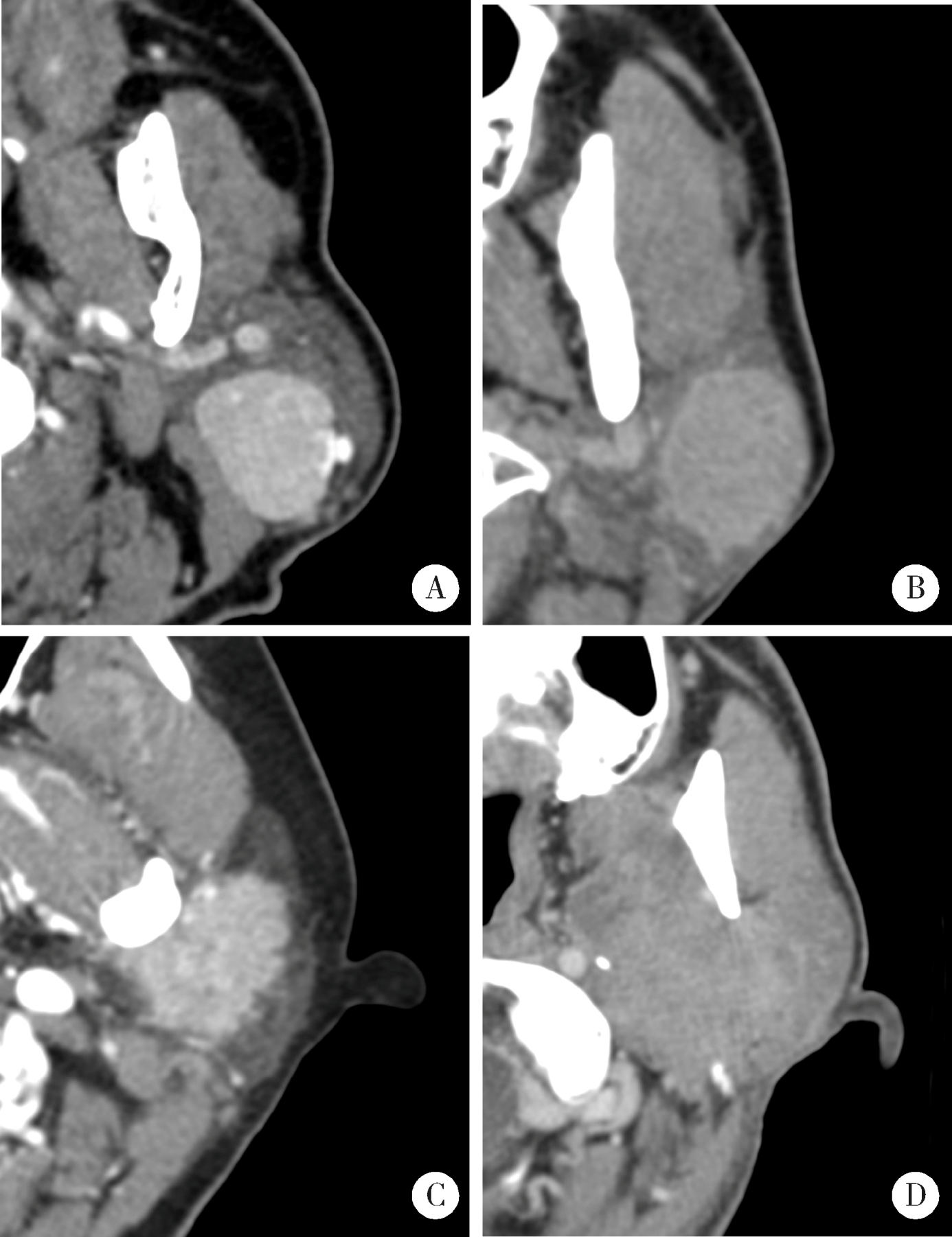
图1

PI-RADS 2级(A)、3级(B)、4级(C)、5级(D)诊断的典型病例"

表2

腮腺CT影像报告与数据系统的诊断标准"

PI-RADS Criteria
1: Normal parotid gland There are definitely no masses in the parotid gland
2: More likely benign (1) Multiple bilateral or unilateral masses presented regular arc border and oval contour with enhancement and without enlarged or necrotic lymph nodes around the parotid gland and neck; (2) A single tumor may also be considered if it is typical (e.g. oval, with obvious and uniform enhancement in the first enhancement phase, with smooth clear border or a low-density fat envelope)
3: Indeterminate (1) Single, round, no enhancement or uniform enhancement, most of the boun-dary clear, for example majorly clear or partially ill-defined; (2) Granular with uneven enhancement but definite boundary; (3) Multiple without enhancement, and without enlarged or necrotic lymph nodes around the parotid gland and neck; (4) Small tumors, that means, all tumors diameter less than 1 cm are included, regardless of their enhancement features
3b: Inflammation including Sjögren
syndrome and IgG4
In multiple gland, lesions are diffuse, with enhancement, without necrotic lymph nodes around the neck
4: Probably malignant (1) Single lesion, the shape is slightly irregular, part of the boundary is indistinctly (the boun-dary marked 3 or 4) or the membrane enhancement with villous edges; (2) The enhancement type is medium enhancement even slightly enhancement, solid lesion with little or without cystic degeneration; (3) Soft tissue density or with partial fluid especially with calcification, a small amount of unilateral diffuse; (4) Have large masses of irregular calcification with swelling lymph nodes but without necrosis lymph nodes
5: Highly suggestive malignancy (1) Necrotic lymph nodes are seen in the neck; (2) The boundary is unclear, the shape is irregular; (3) The border of the surrounding muscles, bones or fat tissues are not clear, which means destruction
6: Already had malignant diagnosis Had pathological result to pronounced it is a malignant neoplasm
7: Unsatisfied illustration on CT The density of neoplasm and the parotid is too nearly to find out what the neoplasm is really like. But couldn’t exclude that a neoplasm may exist

表3

腮腺肿物病理诊断与PI-RADS诊断结果的相关性"

Items PI-RADS 2 PI-RADS 3 PI-RADS 3b PI-RADS 4 PI-RADS 5 PI-RADS 6 PI-RADS total
Warthin tumor 176 72 0 5 0 0 253
Pleomorphic adenoma 4 367 0 23 1 0 395
Basal cell adenoma 11 81 0 2 0 0 94
Myoepithelioma 0 3 0 0 0 0 3
Oncocytoma 4 4 0 0 0 0 8
Cystadenoma 0 6 0 0 0 0 6
Keratocystoma 0 0 0 1 0 0 1
Cyst 28 14 0 0 0 0 42
Lipoma 4 1 0 0 0 0 5
Schwannoma 0 9 0 0 0 0 9
Vascular malformation 2 9 0 2 0 0 13
Eosinophilic lymphogranuloma 0 1 2 0 0 0 3
Inflammation 13 31 9 12 0 0 65
Calcified epithelioma 1 1 0 0 0 0 2
Non-sebaceous lymphoadenoma 0 0 0 1 0 0 1
Lymphoepithelial lesions 0 0 0 3 0 0 3
Nodular fasciitis 0 1 0 0 0 0 1
Myofibromatosis 0 1 0 0 0 0 1
Mucoepidermoid carcinoma 1 6 0 6 6 1 20
Adenocarcinoma, NOS 0 2 0 3 4 0 9
Acinic cell carcinoma 0 13 0 2 0 0 15
Salivary duct carcinoma 0 0 0 1 3 0 4
Adenoid cystic carcinoma 0 4 0 0 3 0 7
Polymorphous adenocarcinoma 0 3 0 5 1 0 9
Uncertainty adenocarcinoma 0 0 1 2 5 0 8
Epithelial-myoepithelial carcinoma 0 1 0 0 0 0 1
Clear cell carcinoma 0 0 0 0 1 0 1
Oncocytic carcinoma 0 0 0 2 0 0 2
Sarcoma 0 1 0 0 0 1 2
Lymphoepithelial carcinoma 0 1 0 2 2 2 7
Malignant lymphoma 0 4 0 3 4 0 11
Malignant melanoma 0 0 0 1 0 0 1
Metastatic solitary fibroma 0 1 0 0 0 0 1
Total 244 637 12 76 30 4 1 003
[1] 张震康, 俞光岩 . 口腔颌面外科学[M]. 2版. 北京: 北京大学医学出版社, 2013.
[2] Abdullah A, Rivas FF, Srinivasan A . Imaging of the salivary glands[J]. Semin Roentgenol, 2013,48(1):65-74.
[3] 俞光岩, 高岩, 孙勇刚 . 口腔颌面部肿瘤[M]. 北京: 人民卫生出版社, 2002.
[4] Seethala RR, Stenman G . Update from the 4th edition of the World Health Organization classification of head and neck tumours: Tumors of the salivary gland[J]. Head Neck Pathol, 2017,11(1):55-67.
[5] White SC, Pharoah MJ . Oral radiology principles and interpretation[M]. 3rd ed. St. Louis, Missouri: Mosby Elsevier, 2014.
[6] 马绪臣 . 口腔颌面医学影像学[M]. 北京: 北京大学医学出版社, 2014.
[7] 马绪臣, 李铁军 . 口腔颌面部疾病CT诊断与鉴别诊断[M]. 北京: 北京大学医学出版社, 2019.
[8] Horvath E, Majlis S, Rossi R , et al. An ultrasonogram reporting system for thyroid nodules stratifying cancer risk for clinical ma-nagement[J]. J Clin Endocrinol Metab, 2009,94(5):1748-1751.
[9] Barentsz JO, Richenberg J, Clements R , et al. ESUR prostate MR guidelines 2012[J]. Eur Radiol, 2012,22(4):746-757.
[10] Abdel Razek AA, Ashmalla GA, Gaballa G , et al. Pilot study of ultrasound parotid imaging reporting and data system (PIRADS): Inter-observer agreement[J]. Eur J Radiol, 2015,84(12):2533-2538.
[11] 马大权, 俞光岩 . 唾液腺病学[M]. 2版. 北京: 人民卫生出版社, 2014.
[12] Kim H, Kim SY, Kim YJ , et al. Correlation between computed tomography imaging and histopathology in pleomorphic adenoma of parotid gland[J]. Auris Nasus Larynx, 2018,45(4):783-790.
[13] Ito FA, Jorge J, Vargas PA , et al. Histopathological findings of pleomorphic adenomas of the salivary glands[J]. Med Oral Patol Oral Cir Bucal, 2009,14(2):E57-61.
[14] 黄敏娴, 马大权, 俞光岩 , 等. 复发性涎腺多形性腺瘤的临床与病理分析[J]. 现代口腔医学杂志, 2008,22(1):1-4.
[1] 季加孚, 韦静涛, 季科, 步召德. 胃癌诊疗的瓶颈与破局:迈向精准化与智能化融合的新纪元[J]. 北京大学学报(医学版), 2026, 58(2): 231-238.
[2] 王海, 江一舟. 靶向血管治疗在乳腺癌精准治疗中的分子机制与临床应用[J]. 北京大学学报(医学版), 2026, 58(2): 251-256.
[3] 付浩, 申潞艳, 黄冰洋, 马少华. 免疫治疗背景下食管鳞状细胞癌围手术期治疗的临床思考[J]. 北京大学学报(医学版), 2026, 58(2): 266-271.
[4] 刘友东, 吕亚军, 陈杰, 臧明德, 潘宏达, 刘晓文, 陆俊, 刘凤林. 全腹腔镜保留贲门胃底胃次全切除术治疗中上部胃癌的疗效及安全性[J]. 北京大学学报(医学版), 2026, 58(2): 301-306.
[5] 李伟浩, 张学民, 李伟, 张韬, 张小明. 胸主动脉腔内修复术左肱动脉穿刺点使用血管缝合器处理的临床效果[J]. 北京大学学报(医学版), 2026, 58(2): 388-392.
[6] 李嘉临, 陈力侨, 唐家天, 吴艳, 王安强. 胃肝样腺癌转化治疗1例[J]. 北京大学学报(医学版), 2026, 58(2): 399-404.
[7] FarinEbrahimi, 冯志强, FarazEbrahimi, 韩玮华, 于子杨, 贾宽宽, 安金刚. 上颌药物相关性颌骨坏死的不同分期手术治疗效果[J]. 北京大学学报(医学版), 2026, 58(1): 107-114.
[8] 赵业, 刁小莉, 熊焰. 细胞转移技术在微量细胞液病理诊断中的应用[J]. 北京大学学报(医学版), 2026, 58(1): 208-213.
[9] 臧海玲, 梁宇红. 上颌第二磨牙慢性根尖周炎合并器械分离的根管再治疗1例[J]. 北京大学学报(医学版), 2026, 58(1): 214-219.
[10] 王月, 梁宇红. 繁茂型牙骨质-骨结构不良1例[J]. 北京大学学报(医学版), 2026, 58(1): 220-224.
[11] 潘莲菲, 李文静, 王瑞洋, 焦剑, 曹战强, 高丽, 释栋. 口服抗生素辅助牙周机械治疗对重度牙周炎的短期疗效及影响因素[J]. 北京大学学报(医学版), 2026, 58(1): 30-36.
[12] 池彦廷, 蒋鸿杰, 陈艳, 徐志秀, 李斌斌. 直接免疫荧光在口腔黏膜寻常型天疱疮诊断中的价值: 基于多指标联合分析的回顾性研究[J]. 北京大学学报(医学版), 2026, 58(1): 68-73.
[13] 顾静妍, 李欣艺, 赵金霞, 穆荣. 误诊为类风湿关节炎、痛风的糖尿病致Charcot关节病1例[J]. 北京大学学报(医学版), 2025, 57(6): 1193-1197.
[14] 肖晓笛, 夏有辰, 柳剑英, 付鹏. 左侧胸锁乳突肌间血管内乳头状内皮增生1例[J]. 北京大学学报(医学版), 2025, 57(5): 1002-1004.
[15] 谢尚, 王鹿鸣, 张馨元, 冯秋实, 夏洋洋, 戴姿薇, 单小峰, 蔡志刚. 口腔鳞癌类器官库的构建及应用[J]. 北京大学学报(医学版), 2025, 57(5): 847-851.
Viewed
Full text


Abstract

Cited

  Shared   
  Discussed   
No Suggested Reading articles found!