北京大学学报(医学版) ›› 2025, Vol. 57 ›› Issue (6): 1165-1173. doi: 10.19723/j.issn.1671-167X.2025.06.022

• 论著 • 上一篇    下一篇

多孔表面结构对立体光固化成型氧化锆疲劳强度的影响

赵健霄1, 丁茜1, 李文锦1, 马全诠1, 兰一笑2, 张磊1,*(), 韩建民2,*()   

  1. 1. 北京大学口腔医学院·口腔医院修复科,北京 100081
    2. 北京大学口腔医学院·口腔医院材料研究室,国家口腔医学中心,国家口腔疾病临床医学研究中心,口腔生物材料和数字诊疗装备国家工程研究中心,国家药品监督管理局口腔材料重点实验室,北京 100081
  • 收稿日期:2023-04-07 出版日期:2025-12-18 发布日期:2024-02-05
  • 通讯作者: 张磊, 韩建民
  • 基金资助:
    北京市自然科学基金(7192233)

Effect of porous surface structure on fatigue strength of 3D printed zirconia

Jianxiao ZHAO1, Qian DING1, Wenjin LI1, Quanquan MA1, Yixiao LAN2, Lei ZHANG1,*(), Jianmin HAN2,*()   

  1. 1. Department of Prosthodontics, Peking University School and Hospital of Stomatology, Beijing 100081, China
    2. Department of Dental Materials, Peking University School and Hospital of Stomatology & National Center for Stomatology & National Clinical Research Center for Oral Diseases & National Engineering Research Center of Oral Biomaterials and Digital Medical Devices & NMPA Key Laboratory for Dental Materials, Beijing 100081, China
  • Received:2023-04-07 Online:2025-12-18 Published:2024-02-05
  • Contact: Lei ZHANG, Jianmin HAN
  • Supported by:
    the Beijing Natural Science Foundation(7192233)

RICH HTML


摘要:

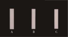
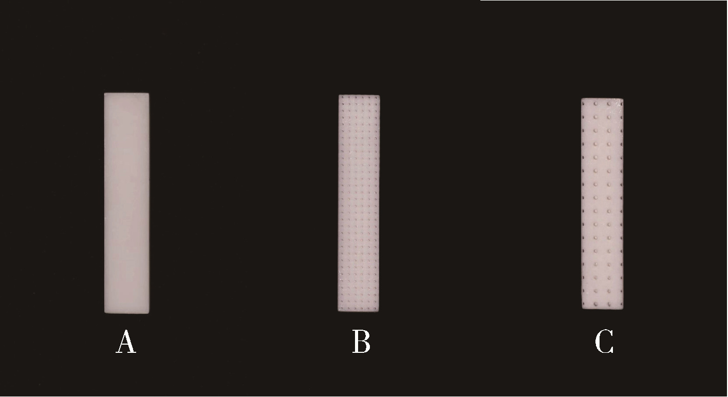
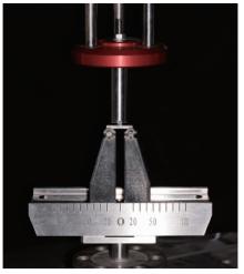
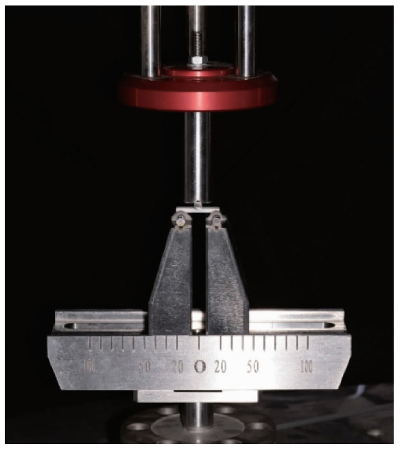
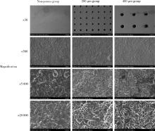
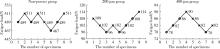
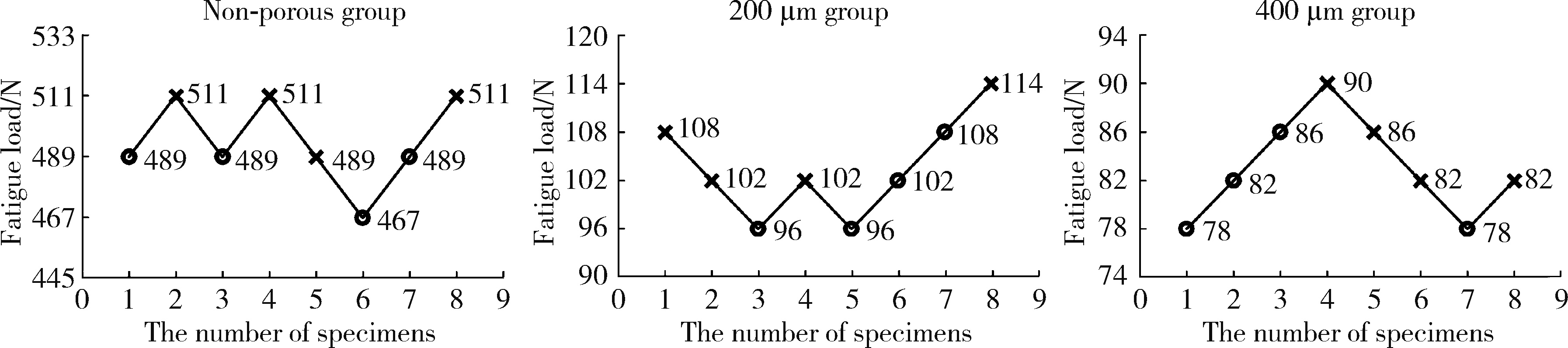
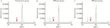
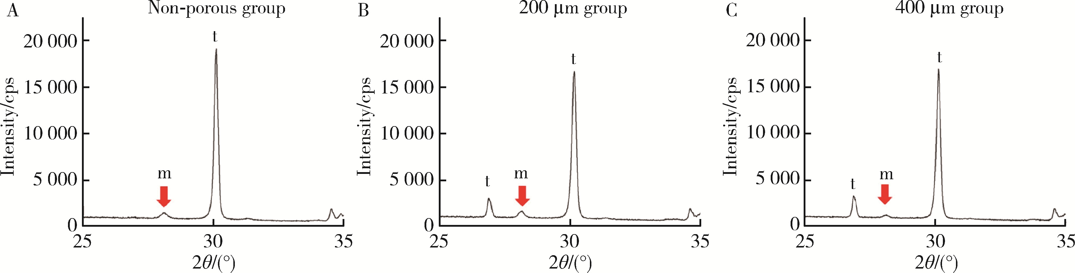
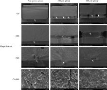
目的: 研究多孔表面结构对立体光固化成型氧化锆疲劳强度的影响,为3D打印氧化锆种植体表面优化设计提供参考。方法: 通过立体光固化成型技术制备氧化锆试件,根据表面结构分为无孔组、200 μm孔组、400 μm孔组。通过三维激光形貌显微镜及扫描电镜观察其表面微观形貌,测定表面粗糙度、孔隙参数、晶粒尺寸。通过三点弯曲试验检测试件弯曲强度,并进行Weibull分析;通过疲劳试验检测试件的疲劳强度,扫描电镜观察断口,采用X射线衍射仪对疲劳试验前后的试件进行晶相分析,分析疲劳机制。结果: 立体光固化成型无孔组、200 μm孔组及400 μm孔组试件表面孔间粗糙度分别为(0.79±0.09) μm、(0.81±0.16) μm、(0.81±0.09) μm,组间差异无统计学意义;表面晶粒尺寸分别为(324.11±21.38) nm、(308.06±11.34) nm、(311.62±15.02) nm,组间差异无统计学意义。三点弯曲试验结果显示,无孔组三点弯曲强度[(1 030.70±111.71) MPa]显著高于两组多孔组(P<0.001),200 μm孔组[(272.04±61.16) MPa]显著高于400 μm孔组[(201.21±25.58) MPa],P<0.01。疲劳试验结果显示,无孔组疲劳强度[(702.29±21.62) MPa]显著高于两组多孔组(P<0.001),200 μm孔组[(159.57±9.30) MPa]显著高于400 μm孔组[(125.36±6.11) MPa],P<0.001。断口分析结果显示,疲劳裂纹源主要为材料内部缺陷、气孔、夹杂及打印层结合处等。疲劳试验前后各组试件之间相比,氧化锆单斜相含量差异均无统计学意义。结论: 表面多孔微观结构会显著降低立体光固化成型氧化锆试件的疲劳强度,且孔径增大可造成疲劳强度下降;未来应着重改进3D打印氧化锆的材料及打印工艺,进一步优化表面结构设计,以提升3D打印氧化锆的力学性能。

关键词: 氧化锆, 多孔, 3D打印, 疲劳强度

Abstract:

Objective: To study the effect of porous surface structure on fatigue strength of zirconia fabricated by stereolithography apparatus (SLA), and to provide reference for the surface design of 3D printed zirconia implants. Methods: Zirconia specimens were fabricated by SLA. According to the surface structure, zirconia specimens were divided into non-porous group, 200 μm group and 400 μm group. The surface morphology was observed by 3D laser morphology microscope and scanning electron microscope, and the surface roughness, pore parameters and grain size were measured. The flexural strength of the specimen was measured by three-point bending test and Weibull analysis was performed. The fatigue strength of the specimens was measured by fatigue test, and the fatigue mechanism was analyzed by fractrography. The crystal phase before and after fatigue test of the specimen was analyzed by X-ray diffraction. Results: The surface roughness of the area between the pores of non-porous group, 200 μm group and 400 μm group was (0.79±0.09) μm, (0.81±0.16) μm and (0.81±0.09) μm, respectively, with no significant difference among them. The surface grain size was (324.11±21.38) nm, (308.06±11.34) nm, (311.62±15.02) nm, respectively, with no significant difference among them. The results of three-point bending test showed that the three-point bending strength of the non-porous group [(1 030.70±111.71) MPa] was significantly higher than that of the porous groups (P < 0.001). The 200 μm group [(272.04±61.16) MPa] was significantly higher than the 400 μm group [(201.21±25.58) MPa] (P < 0.01). The fatigue strength of the non-porous group [(702.29± 21.62) MPa] was significantly higher than that of the porous groups (P < 0.001), and the fatigue strength of the 200 μm group [(159.57±9.30) MPa] was significantly higher than that of the 400 μm group [(125.36±6.11) MPa] (P < 0.001). The fracture analysis results showed that the crack origins were mainly internal defects, air holes, inclusions and the joint of printing layer, etc. There was no significant difference in the content of monoclinic phase before and after fatigue test among all the groups. Conclusion: The surface porous microstructure could significantly reduce the fatigue strength of the zirconia specimens, and the larger pore size showed the lower fatigue strength. In the future, the material and printing process of 3D printing zirconia should be improved, and the surface structure design should be further optimized to improve the mechanical properties of 3D printing zirconia.

Key words: Zirconia, Porous, 3D printing, Fatigue strength

中图分类号: 

  • R783.1

图1

氧化锆试件设计制备流程"

图2

3D打印氧化锆试件烧结曲线"

图3

各组3D打印氧化锆试件"

图4

立体光固化成型氧化锆试件疲劳强度试验"

表1

氧化锆试件孔间粗糙度"

Group Ra/μm Rq/μm Rz/μm
Non-porous 0.79±0.09 0.98±0.21 8.17±1.02
200 μm 0.81±0.16 1.06±0.18 8.79±1.48
400 μm 0.81±0.09 1.05±0.11 8.31±0.89

表2

多孔氧化锆试件表面孔隙参数测量"

Group Design value of
pore diameter/μm
Measured value of
pore diameter/μm, ${\bar x}$±s
Design value of
pore depth/μm
Measured value of
pore depth/μm, ${\bar x}$±s
Design value of
pore-pitch/μm
Measured value of
pore-pitch/μm,${\bar x}$±s
200 μm 200.00 200.02±6.15 400.00 379.35±11.22** 430.00 429.08±9.31
400 μm 400.00 366.48±6.88** 600.00 572.84±29.35* 860.00 883.99±9.41**

图5

氧化锆试件表面扫描电镜观察"

表3

氧化锆试件表面晶粒尺寸"

Group Average grain size/nm, ${\bar x}$±s
Non-porous 324.11±21.38
200 μm 308.06±11.34
400 μm 311.62±15.02

表4

氧化锆试件三点弯曲强度及疲劳强度"

Group Three-point flexural
strength/MPa, ${\bar x}$±s
Fatigue strength/MPa,
${\bar x}$±s
Characteristic
strength/MPa
95%CI Weibull modulus 95%CI
Non-porous 1 030.70±111.71 702.29±21.62 1 080.10 1 027.12-1 135.91 10.61 7.23-15.84
200 μm 272.04±61.16 159.57±9.30 296.40 264.97-331.55 4.80 3.29-6.99
400 μm 201.21±25.58 125.36±6.11 211.68 201.25-222.66 10.50 6.89-15.99

图6

升降法疲劳试验结果"

表5

氧化锆试件表面单斜晶相比例"

Group Before fatigue test Unbroken after fatigue test Broken after fatigue test
Xm Vm Xm Vm Xm Vm
Non-porous 9.51% 12.09% 10.78% 13.67% 10.42% 13.23%
200 μm 10.01% 12.72% 11.17% 14.14% 10.43% 13.25%
400 μm 9.64% 12.27% 9.69% 12.34% 10.42% 13.22%

图7

氧化锆试件(疲劳试验前)X射线衍射图谱,m相为单斜相氧化锆波峰,t相为四方相氧化锆波峰"

图8

氧化锆试件疲劳破坏断口扫描电镜观察"

1
赵祯, 代康, 高勃. 3D打印陶瓷技术在口腔医学领域的研究进展[J]. 中国实用口腔科杂志, 2021, 14 (6): 739- 744.
2
Osman RB , van der Veen AJ , Huiberts D , et al. 3D-printing zirconia implants; a dream or a reality? An in-vitro study evaluating the dimensional accuracy, surface topography and mechanical properties of printed zirconia implant and discs[J]. J Mech Behav Biomed Mater, 2017, 75, 521- 528.

doi: 10.1016/j.jmbbm.2017.08.018
3
Thakur J , Parlani S , Shivakumar S , et al. Accuracy of marginal fit of an implant-supported framework fabricated by 3D printing versus subtractive manufacturing technique: A systematic review and meta-analysis[J]. J Prosthet Dent, 2023, 129 (2): 301- 309.

doi: 10.1016/j.prosdent.2021.05.010
4
Sakthiabirami K , Kang JH , Jang JG , et al. Hybrid porous zirconia scaffolds fabricated using additive manufacturing for bone tissue engineering applications[J]. Mater Sci Eng C Mater Biol Appl, 2021, 123, 111950.

doi: 10.1016/j.msec.2021.111950
5
中国国家标准化管理委员会. 金属平均晶粒度测定方法: GB/T 6394—2017[J]. 北京: 中国标准出版社, 2017, 8- 13.
6
International Organization for Standardization . Dentisry-ceramic materials: ISO 6872: 2015[J]. Switzerland: ISO, 2015, 7- 16.
7
Studart AR , Filser F , Kocher P , et al. Fatigue of zirconia under cyclic loading in water and its implications for the design of dental bridges[J]. Dent Mater, 2007, 23 (1): 106- 114.

doi: 10.1016/j.dental.2005.12.008
8
Zucuni CP , Guilardi LF , Rippe MP , et al. Fatigue strength of yttria-stabilized zirconia polycrystals: Effects of grinding, poli-shing, glazing, and heat treatment[J]. J Mech Behav Biomed Mater, 2017, 75, 512- 520.

doi: 10.1016/j.jmbbm.2017.06.016
9
Ding Q , Zhang L , Bao R , et al. Effects of different surface treatments on the cyclic fatigue strength of one-piece CAD/CAM zirconia implants[J]. J Mech Behav Biomed Mater, 2018, 84, 249- 257.

doi: 10.1016/j.jmbbm.2018.05.002
10
Dehurtevent M , Robberecht L , Hornez JC , et al. Stereolithography: A new method for processing dental ceramics by additive computer-aided manufacturing[J]. Dent Mater, 2017, 33 (5): 477- 485.

doi: 10.1016/j.dental.2017.01.018
11
Zhai Z , Sun J . Research on the low-temperature degradation of dental zirconia ceramics fabricated by stereolithography[J]. J Prosthet Dent, 2023, 130 (4): 629- 638.

doi: 10.1016/j.prosdent.2021.11.012
12
Kohal RJ , Bächle M , Att W , et al. Osteoblast and bone tissue response to surface modified zirconia and titanium implant materials[J]. Dent Mater, 2013, 29 (7): 763- 776.

doi: 10.1016/j.dental.2013.04.003
13
Al Qahtani WMS , Schille C , Spintzyk S , et al. Effect of surface modification of zirconia on cell adhesion, metabolic activity and proliferation of human osteoblasts[J]. Biomed Tech (Berl), 2017, 62 (1): 75- 87.
14
Hafezeqoran A , Koodaryan R . Effect of zirconia dental implant surfaces on bone integration: A systematic review and meta-analysis[J]. Biomed Res Int, 2017, 2017, 9246721.
15
Boyan BD , Hummert TW , Dean DD , et al. Role of material surfaces in regulating bone and cartilage cell response[J]. Biomaterials, 1996, 17 (2): 137- 146.

doi: 10.1016/0142-9612(96)85758-9
16
Hadjicharalambous C , Buyakov A , Buyakova S , et al. Porous alumina, zirconia and alumina/zirconia for bone repair: Fabrication, mechanical and in vitro biological response[J]. Biomed Mater, 2015, 10 (2): 025012.

doi: 10.1088/1748-6041/10/2/025012
17
Cheng A , Humayun A , Cohen DJ , et al. Additively manufactured 3D porous Ti-6Al-4V constructs mimic trabecular bone structure and regulate osteoblast proliferation, differentiation and local factor production in a porosity and surface roughness dependent manner[J]. Biofabrication, 2014, 6 (4): 045007.

doi: 10.1088/1758-5082/6/4/045007
18
Palmero P . Structural ceramic nanocomposites: A review of pro-perties and powders' synthesis methods[J]. Nanomaterials (Basel), 2015, 5 (2): 656- 696.

doi: 10.3390/nano5020656
19
Dos-Santos C , Santos FA , Elias CN . Properties of NAnostructured 3Y-TZP blocks used for CAD/CAM dental restoration[J]. Key Eng Mater, 2008, 396/397/398, 603- 606.
20
Silva CP , Santos C , Silva CRM . Mechanical properties of nanostructured zirconia[J]. Mater Sci Forum, 2010, 660/661, 757- 761.

doi: 10.4028/www.scientific.net/MSF.660-661.757
21
Egilmez F , Ergun G , Cekic-Nagas I , et al. Factors affecting the mechanical behavior of Y-TZP[J]. J Mech Behav Biomed Mater, 2014, 37, 78- 87.

doi: 10.1016/j.jmbbm.2014.05.013
22
Gonzaga CC , Cesar PF , Miranda WG Jr , et al. Slow crack growth and reliability of dental ceramics[J]. Dent Mater, 2011, 27 (4): 394- 406.

doi: 10.1016/j.dental.2010.10.025
23
Lakhdar Y , Tuck C , Binner J , et al. Additive manufacturing of advanced ceramic materials[J]. Prog Mater Sci, 2021, 116, 100736.

doi: 10.1016/j.pmatsci.2020.100736
24
李文利, 周宏志, 刘卫卫, 等. 光固化3D打印陶瓷浆料及流变性研究进展[J]. 材料工程, 2022, 50 (7): 40- 50.
25
Karakoca S , Yilmaz H . Influence of surface treatments on surface roughness, phase transformation, and biaxial flexural strength of Y-TZP ceramics[J]. J Biomed Mater Res B Appl Biomater, 2009, 91 (2): 930- 937.
26
Conti L , Bienenstein D , Borlaf M , et al. Effects of the layer height and exposure energy on the lateral resolution of zirconia parts printed by lithography-based additive manufacturing[J]. Materials (Basel), 2020, 13 (6): 1317.

doi: 10.3390/ma13061317
27
Wang B , Arab A , Xie J , et al. The influence of microstructure on the flexural properties of 3D printed zirconia part via digital light processing technology[J]. Materials (Basel), 2022, 15 (4): 1602.

doi: 10.3390/ma15041602
[1] 钱锟, 刘亦洪. 基于直接法和间接法数字印模制作的高嵌体适合性评价的体外研究[J]. 北京大学学报(医学版), 2025, 57(3): 604-609.
[2] 闵树元, 田耘. 3D打印生物可降解WE43镁合金支架的生物相容性及对骨缺损的治疗[J]. 北京大学学报(医学版), 2025, 57(2): 309-316.
[3] 展新新,曹露露,项东,汤皓,夏丹丹,林红. 成型方向对3D打印口腔义齿基托树脂材料物理性能及力学性能的影响[J]. 北京大学学报(医学版), 2024, 56(2): 345-351.
[4] 胡攀攀,李彦,刘啸,唐彦超,李梓赫,刘忠军. 自稳式人工椎体在颈椎前路手术中的应用[J]. 北京大学学报(医学版), 2024, 56(1): 161-166.
[5] 丁茜,李文锦,孙丰博,谷景华,林元华,张磊. 表面处理对氧化钇和氧化镁稳定的氧化锆种植体晶相及断裂强度的影响[J]. 北京大学学报(医学版), 2023, 55(4): 721-728.
[6] 周华,王仁吉,刘忠军,刘晓光,吴奉梁,党礌,韦峰. 3D打印人工椎体在颈椎脊索瘤全脊椎切除术中的应用[J]. 北京大学学报(医学版), 2023, 55(1): 144-148.
[7] 李伟伟,陈虎,王勇,孙玉春. 氧化锆陶瓷表面硅锂喷涂层的摩擦磨损性能[J]. 北京大学学报(医学版), 2023, 55(1): 94-100.
[8] 开地尔娅·阿不都热合曼,张荣赓,钱浩楠,邹振洋,丹尼娅·叶斯涛,范田园. 个性化剂量熔融沉积成型3D打印茶碱片剂的制备和体外评价[J]. 北京大学学报(医学版), 2022, 54(6): 1202-1207.
[9] 邓艺,张一,李博文,王梅,唐琳,刘玉华. 不同交联剂处理对脱细胞小肠黏膜下层多孔支架的影响[J]. 北京大学学报(医学版), 2022, 54(3): 557-564.
[10] 王铮,丁茜,高远,马全诠,张磊,葛兮源,孙玉春,谢秋菲. 氧化锆多孔表面显微形貌对成骨细胞增殖及分化的影响[J]. 北京大学学报(医学版), 2022, 54(1): 31-39.
[11] 孙玉春,郭雨晴,陈虎,邓珂慧,李伟伟. 口腔精准仿生修复技术的自主创新研发与转化[J]. 北京大学学报(医学版), 2022, 54(1): 7-12.
[12] 李文锦,丁茜,原福松,孙丰博,郑剑桥,鲍蕊,张磊. 飞秒激光表面处理对氧化锆表面特征及弯曲强度的影响[J]. 北京大学学报(医学版), 2021, 53(4): 770-775.
[13] 杨欣,李榕,叶红强,陈虎,王勇,周永胜,孙玉春. 不同刃状边缘补偿角度的两种氧化锆全瓷冠断裂强度的评价[J]. 北京大学学报(医学版), 2021, 53(2): 402-405.
[14] 郑苗,詹凌璐,刘志强,李和平,谭建国. 不同等离子体处理氧化锆对人牙龈成纤维细胞黏附能力的影响[J]. 北京大学学报(医学版), 2019, 51(2): 315-320.
[15] 周团锋,王新知. 计算机辅助设计与制作的一体化氧化锆全瓷桩核5年临床观察[J]. 北京大学学报(医学版), 2018, 50(4): 680-684.
Viewed
Full text


Abstract

Cited

  Shared   
  Discussed   
No Suggested Reading articles found!