北京大学学报(医学版) ›› 2020, Vol. 52 ›› Issue (5): 924-930. doi: 10.19723/j.issn.1671-167X.2020.05.022
三维图像融合技术评价上颌全牙列种植固定修复前后的鼻唇软组织形态变化
郝柯屹,罗佳,邸萍,郭厚佐,沈惠丹,刘焱萍,张宇( ),林野
),林野
- 北京大学口腔医学院·口腔医院,种植科 国家口腔疾病临床医学研究中心 口腔数字化医疗技术和材料国家工程实验室 口腔数字医学北京市重点实验室,北京 100081
Validation of the digital integration technology for evaluating the nasolabial morphology variation after the cross-arch fixed restoration of maxillary implant-supported prostheses
Ke-yi HAO,Jia LUO,Ping DI,Hou-zuo GUO,Hui-dan SHEN,Yan-ping LIU,Yu ZHANG(),Ye LIN
- Department of Oral Implantology, Peking University School and Hospital of Stomatology & National Clinical Research Center for Oral Diseases & National Engineering Laboratory for Digital and Material Technology of Stomatology & Beijing Key Laboratory of Digital Stomatology, Beijing 100081, China
摘要:
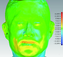
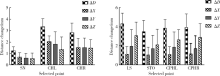
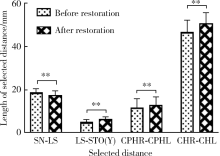
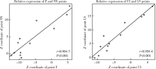
目的:探讨利用面部与义齿三维(three dimensional,3D)图像融合方法评估全牙列种植固定修复前后鼻唇区软组织形态变化的可行性。方法:选择于北京大学口腔医院种植科就诊的上颌无牙颌患者共12例(女性4例、男性8例),平均年龄(54.82±5.50)岁(45~62岁)。于上颌植入4~6枚种植体,6个月后为患者佩戴全牙列固定义齿,捕捉患者佩戴义齿前后的3D面相及义齿形态数据。将三维数据置于同一坐标系中,对义齿和软组织标志点进行测量,分别比较各标志点[左口角点(cheilion left, CHL)、右口角点(cheilion right, CHR)、左唇峰点(crista philtri left,CPHL)、右唇峰点(crista philtri right, CPHR)、上唇缘点(labrale superius, LS)、鼻下点(subnasale, SN)、口裂正中点(stomion,STO)、上中切牙点(upper incisor, UI)、义齿上边缘顶点(F-point, F)]的位移量及标志点间线距[人中长度(SN-LS)、双唇峰间距(CPHR-CPHL)、口裂宽(CHR-CHL)、上唇红高度(LS-STO)]变化。结果:义齿与软组织拟合后,义齿上边缘顶点与鼻下点直线距离(F-SN)三次测量结果的一致性检验显示该方法可重复性好,组内相关系数(intraclass correlation coefficient, ICC)为0.983 (95%CI:0.957~0.995)。佩戴义齿后,软组织各标志点均发生前徙变化,鼻底区域变化最小,SN前后方向上的位移为(0.61±0.44) mm,LS前后向位移为(3.12±1.38) mm。在垂直方向上,SN、LS、CPHL、CPHR均向上移动,STO、CHL、CHR向下方少量移动。除SN-LS略微减小外,CHR-CHL、CPHR-CPHL、LS-STO均增大(P<0.01)。SN与F、LS与UI的Z轴坐标值均呈现高度相关(r=0.904 3、r=0.958 4)。结论:三维面像与义齿图像融合方法可重复性好,经该方法检测,义齿与软组织标志点的前后向位移之间存在高度相关性。
中图分类号:
- R783






| [1] |
Kau CH, Richmond S, Zhurov A, et al. Use of 3-dimensional surface acquisition to study facial morphology in 5 populations[J]. Am J Orthod Dentofacial Orthop, 2010,137(4 Suppl):S56-57.
pmid: 20381762 |
| [2] |
Karatas OH, Toy E. Three-dimensional imaging techniques: A literature review[J]. Eur J Dent, 2014,8(1):132-140.
pmid: 24966761 |
| [3] |
Maló P, de Araújo Nobre M, Lopes A, et al. “All-on-4” imme-diate-function concept for completely edentulous maxillae: a clinical report on the medium (3 years) and long-term (5 years) outcomes[J]. Clin Implant Dent Relat Res, 2012,14(Suppl 1):e139-150.
doi: 10.1111/j.1708-8208.2011.00395.x |
| [4] |
Lopes A, Maló P, de Araújo Nobre M, et al. The NobelGuide® All-on-4® treatment concept for rehabilitation of edentulous jaws: a retrospective report on the 7-years clinical and 5-years radiographic outcomes [J]. Clin Implant Dent Relat Res, 2017,19(2):233-244.
pmid: 27758069 |
| [5] | 张宇, 林野, 刘洋, 等. 牙周炎晚期伴上颌牙槽骨前突畸形患者即刻种植全牙列固定修复的侧貌变化初探[J]. 中华口腔医学杂志, 2017,52(10):625-630. |
| [6] | 邸萍, 林野, 李健慧, 等. 单颌拔牙后即刻种植即刻修复的临床回顾研究[J]. 中华口腔医学杂志, 2013,48(4):216-222. |
| [7] | Holzinger D, Seemann R, Matoni N, et al. Effect of dental implants on bisphosphonate-related osteonecrosis of the jaws[J]. J Oral Maxillofac Surg, 2014, 72(10): 1937.e1-8. |
| [8] |
Tian K, Li Q, Wang X, et al. Reproducibility of natural head position in normal Chinese people[J]. Am J Orthod Dentofacial Orthop, 2015,148(3):503-510.
pmid: 26321348 |
| [9] |
Huang Y, Zhang X, Fan Y, et al. Reshaping 3D facial scans for facial appearance modeling and 3D facial expression analysis[J]. Image and Vision Computing, 2012,30(10):750-761.
doi: 10.1016/j.imavis.2011.12.008 |
| [10] |
Littlefield T, Kelly K, Cherney J, et al. Development of a new three-dimensional cranial imaging system[J]. J Craniofac Surg, 2004,15(1):175-181.
pmid: 14704586 |
| [11] |
Rangel FA, Maal TJ, Bergé SJ, et al. Integration of digital dental casts in 3-dimensional facial photographs[J]. Am J Orthod Dentofacial Orthop, 2008,134(6):820-826.
pmid: 19061810 |
| [12] |
Rosati R, De Menezes M, Rossetti A, et al. Digital dental cast placement in 3-dimensional, full-face reconstruction: a technical evaluation[J]. Am J Orthod Dentofacial Orthop, 2010,138(1):84-88.
pmid: 20620838 |
| [13] |
Avrampou M, Mericske-Stern R, Blatz MB, et al. Virtual implant planning in the edentulous maxilla: criteria for decision making of prosjournal design[J]. Clin Oral Implants Res, 2013,24(Suppl A100):152-159.
doi: 10.1111/j.1600-0501.2011.02407.x |
| [14] |
Mu CQ, Wang SQ, Liu Y, et al. Development of a facescan 3D facial reconstruction technology method for quantitative evaluation of cheilitis granulomatosa[J]. Sci Rep, 2017,7(1):1295.
pmid: 28465526 |
| [15] |
Kamashita Y, Kamada Y, Kawahata N, et al. Influence of lip support on the soft-tissue profile of complete denture wearers[J]. J Oral Rehabil, 2006,33(2):102-109.
doi: 10.1111/j.1365-2842.2006.01575.x pmid: 16457669 |
| [16] |
Fourie Z, Damstra J, Gerrits PO, et al. Evaluation of anthropometric accuracy and reliability using different three-dimensional scanning systems[J]. Forensic Sci Int, 2011,207(1-3):127-134.
doi: 10.1016/j.forsciint.2010.09.018 |
| [17] |
Metzger TE, Kula KS, Eckert GJ, et al. Orthodontic soft-tissue parameters: a comparison of cone-beam computed tomography and the 3dMD imaging system[J]. Am J Orthod Dentofacial Orthop, 2013,144(5):672-681.
pmid: 24182583 |
| [18] |
Desesa CR, Metzler P, Sawh-Martinez R, et al. Three-dimensional nasolabial morphologic alterations following Le Fort I[J]. Plast Reconstr Surg Glob Open, 2016,4(8):e848.
pmid: 27622116 |
| [19] |
Mccollum AGH, Dancaster JT, Evans WG, et al. Sagittal soft-tissue changes related to the surgical correction of maxillary-deficient Class III malocclusions[J]. Seminars in Orthodontics, 2009,15(3):172-184.
doi: 10.1053/j.sodo.2009.03.003 |
| [20] | 卫彦, 陈贵, 韩冰, 等. 三维照相定量评价总义齿修复前后面部软组织变化[J]. 北京大学学报(医学版), 2014,46(1):100-103. |
| [1] | 刘嘉昱, 祝宁, 张育祯, 高贤明, 张宇. 动态导航辅助环钻取骨的准确性[J]. 北京大学学报(医学版), 2026, 58(2): 365-371. |
| [2] | 杨咏涛, 田淯文, 单珅瑶, 李文博, 商相宜, 王艺蓁, 郭殊玮, 高梓翔, 温奥楠, 赵一姣, 王勇. 基于多视图立体视觉的无牙颌种植固定修复软组织数字印模的方法[J]. 北京大学学报(医学版), 2026, 58(1): 126-132. |
| [3] | 于录, 吴灵, 刘筱菁, 李自力. 基于数据库相似性检索的正颌外科手术规划技术流程可行性研究: 随机对照试验[J]. 北京大学学报(医学版), 2026, 58(1): 145-152. |
| [4] | 王宇蓝, 曾浩, 张玉峰. 口腔种植中血浆基质的临床转化现状与前沿探索[J]. 北京大学学报(医学版), 2025, 57(5): 836-840. |
| [5] | 于子杨, 郭厚佐, 蒋析, 韩玮华, 林野. 穿颧种植体上颌窦段成骨的影像学研究[J]. 北京大学学报(医学版), 2025, 57(5): 967-974. |
| [6] | 马丽娟, 腾雍辉, 王勇, 赵一姣, 张馨月, 秦庆钊, 尹东. 乳牙缺失数字化丝圈间隙保持器的三维有限元分析[J]. 北京大学学报(医学版), 2025, 57(2): 376-383. |
| [7] | 方媛媛, 徐帆, 雷杰, 张昊, 张文宇, 孙宇, 吴宏新, 傅开元, 毛伟玉. 基于颞下颌关节紊乱病诊断标准的临床自动诊断系统的建立及验证[J]. 北京大学学报(医学版), 2025, 57(1): 192-201. |
| [8] | 王鹃, 邱立新, 尉华杰. 下颌磨牙穿龈形态设计对种植体周围软组织影响的随机对照临床研究[J]. 北京大学学报(医学版), 2025, 57(1): 65-72. |
| [9] | 李虹, 马斐斐, 翁金龙, 杜阳, 吴彬彰, 孙凤. 口腔即刻种植时动态导航系统的种植精度分析[J]. 北京大学学报(医学版), 2025, 57(1): 85-90. |
| [10] | 徐心雨,吴灵,宋凤岐,李自力,张益,刘筱菁. 基于下颌运动轨迹的正颌外科术中下颌骨髁突定位方法及初步精度验证[J]. 北京大学学报(医学版), 2024, 56(1): 57-65. |
| [11] | 王聪伟,高敏,于尧,章文博,彭歆. 游离腓骨瓣修复下颌骨缺损术后义齿修复的临床分析[J]. 北京大学学报(医学版), 2024, 56(1): 66-73. |
| [12] | 李穗,马雯洁,王时敏,丁茜,孙瑶,张磊. 上前牙种植单冠修复体切导的数字化设计正确度[J]. 北京大学学报(医学版), 2024, 56(1): 81-87. |
| [13] | 刘晓强,周寅. 牙种植同期植骨术围术期高血压的相关危险因素[J]. 北京大学学报(医学版), 2024, 56(1): 93-98. |
| [14] | 丁茜,李文锦,孙丰博,谷景华,林元华,张磊. 表面处理对氧化钇和氧化镁稳定的氧化锆种植体晶相及断裂强度的影响[J]. 北京大学学报(医学版), 2023, 55(4): 721-728. |
| [15] | 欧蒙恩,丁云,唐卫峰,周永胜. 基台边缘-牙冠的平台转移结构中粘接剂流动的三维有限元分析[J]. 北京大学学报(医学版), 2023, 55(3): 548-552. |
|
||
