北京大学学报(医学版) ›› 2020, Vol. 52 ›› Issue (1): 107-112. doi: 10.19723/j.issn.1671-167X.2020.01.017
无牙颌患者鼻唇角变化侧面观的视觉敏感阈值
游浪1,邓珂慧2,李伟伟2,赵一姣2,△( ),孙玉春2,△(),周永胜1
- 1. 北京大学口腔医学院·口腔医院, 修复科, 北京 100081
2. 北京大学口腔医学院·口腔医院,口腔医学数字化研究中心 国家口腔疾病临床医学研究中心 口腔数字化医疗技术和材料国家工程实验室 口腔数字医学北京市重点实验室,北京 100081
Visual sensitivity threshold of lateral view of nasolabial Angle changes in edentulous jaw patients
Lang YOU1,Ke-hui DENG2,Wei-wei LI2,Yi-jiao ZHAO2,△(),Yu-chun SUN2,△(),Yong-sheng ZHOU1
- 1. Center of Digital Dentistry, Peking University School and Hospital of Stomatology & Department of Prosthodontics, Beijing 100081, China
2. Center of Digital Dentistry, Peking University School and Hospital of Stomatology & National Clinical Research Center for Oral Diseases & National Engineering Laboratory for Digital and Material Technology of Stomatology & Beijing Key Laboratory of Digital Stomatology, Beijing 100081, China
摘要:
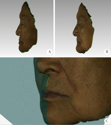
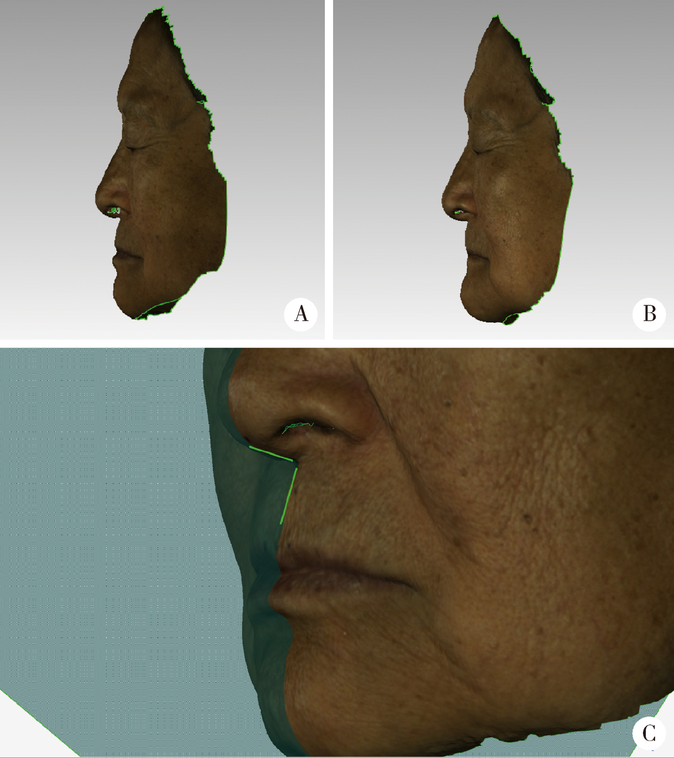
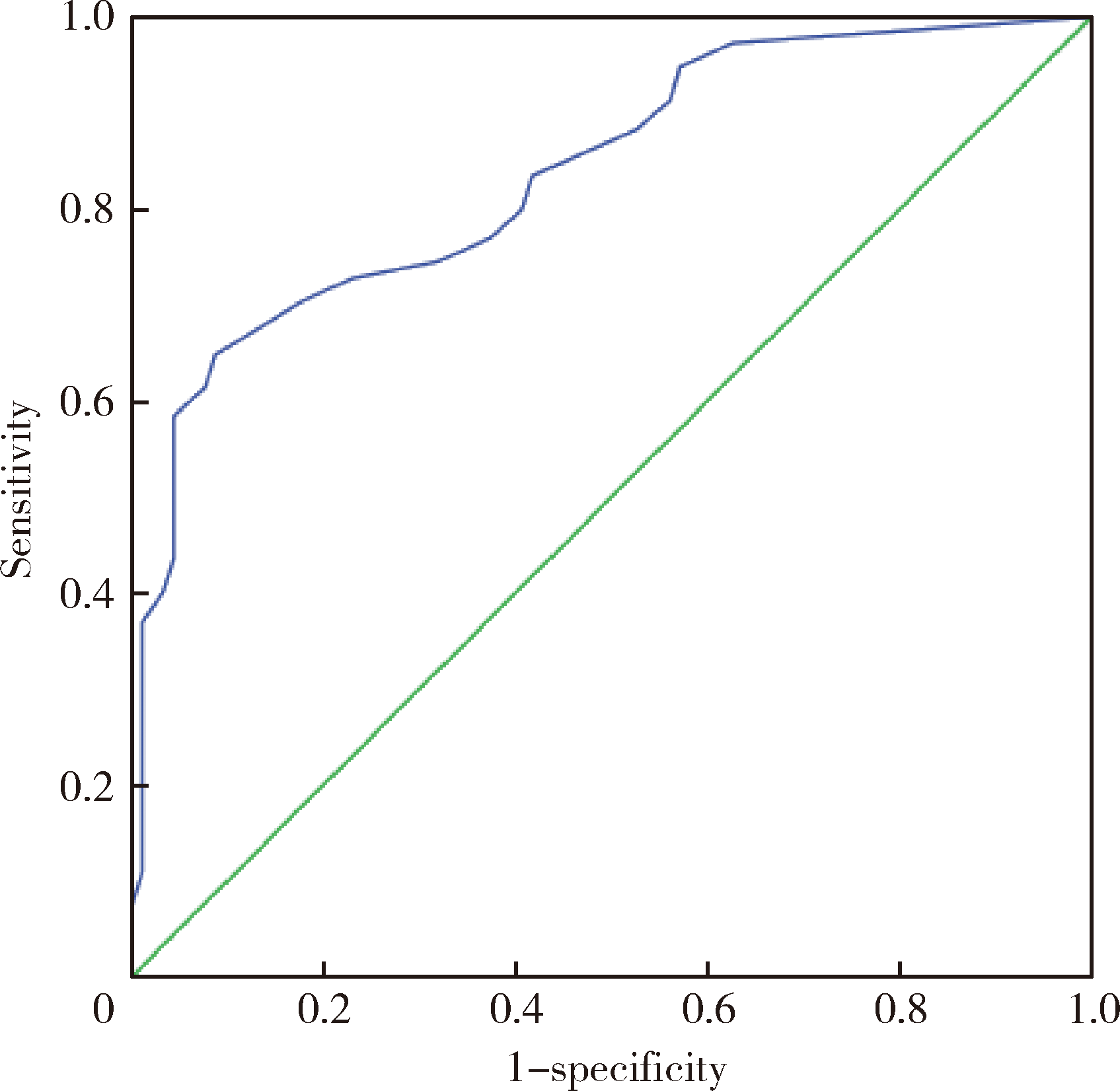
目的:研究医师肉眼对无牙颌患者鼻唇角差异侧面观的视觉敏感阈值,明确人眼可有效识别的鼻唇角差异大小,以期为软组织侧貌审美评价相关研究提供人眼可识别的鼻唇角差异的参考值。方法:获取3名无牙颌患者戴入不同唇部支撑诊断义齿的面部三维数据,对患者的面部三维数据进行同比例侧面截图,以口腔修复科医师(主任医师)和患者肉眼观察双重确认的合适唇丰满度时的侧面截图为参照图片,将其他侧面截图分别与之组合成组,由受试者以随机顺序观察。选择15名口腔医师作为受试者观察每组图片的鼻唇角差异情况,测量计算每组两张图片的鼻唇角差值。实验结果绘制成受试者工作特征(ROC)曲线,使用SPSS 20.0统计软件及约登(Youden)指数计算最佳截断值作为医师肉眼对无牙颌患者鼻唇角差异侧面观的视觉敏感阈值。结果:15名受试者数据分别绘制ROC曲线,其中最佳截断值最大者为5.55°,最小者为3.12°。数据汇总后绘制整体受试者工作特征曲线,其最佳截断值为5.36°(AUC=0.84>0.5, P=0.000<0.05)。当图片的鼻唇角差异在5.36°以上时,受试者能够有效识别。结论:人对鼻唇角变化量的视觉感知存在极限,研究得出了5.36°的鼻唇角差异侧面观的视觉敏感阈值。低于此值的鼻唇角差异可能无临床意义,此结果为软组织侧貌审美评价提供了人眼可识别的鼻唇角变化量阈值的参考值,可应用于软组织侧貌的美学评估,以及作为以鼻唇角为指标进行精度评价的相关研究的误差等级。
中图分类号:
- R783


| [1] | Harris R, Nagarkar P, Amirlak B . Varied definitions of nasolabial angle[J]. Plast Reconstr Surg Glob Open, 2016,4(6):e752. |
| [2] | 徐安秀, 邓锋, 王芬芬 , 等. 鼻唇角改变对骨性Ⅰ类软组织侧貌影响的审美评价[J]. 华西口腔医学杂志, 2015,33(5):492-496. |
| [3] | 马玥, 任嫒姝, 付钢 , 等. 鼻唇角、颏唇角改变对骨性Ⅱ类和Ⅲ类患者面容影响的三维美学评价[J]. 实用口腔医学杂志, 2017,33(5):647-652. |
| [4] | Desesa CR, Metzler P, Sawh-Martinez R , et al. Three-dimensional nasolabial morphologic alterations following Le Fort I[J]. Plast Reconstr Surg Glob Open, 2016,4(8):e848. |
| [5] | Aniruddh YV, Ravi K, Edeinton A. Comparative evaluation of soft tissue changes in Class Ⅰ borderline patients treated with extraction and nonextraction modalities[J]. Dental Press J Orthod, 2016,21(4):50-59. |
| [6] | Kamashita Y, Kamada Y, Kawahata N , et al. Influence of lip support on the soft-tissue profile of complete denture wearers[J]. J Oral Rehabil, 2006,33(2):102-109. |
| [7] | Kaipatur NR, Flores-Mir C . Accuracy of computer programs in predicting orthognathic surgery soft tissue response[J]. J Oral Maxillofac Surg, 2009,67(4):751-759. |
| [8] | Raschke GF, Rieger UM, Bader RD , et al. Perioral aging: an anthropometric appraisal[J]. J Craniomaxillofac Surg, 2014,42(5):e312-e317. |
| [9] | Sierpinska T, Golebiewska M, Kuc J , et al. The influence of the occlusal vertical dimension on masticatory muscle activities and hyoid bone position in complete denture wearers[J]. Adv Med Sci, 2009,54(1):104-108. |
| [10] | Krajicek DD . Guides for natural facial appearance as related to complete denture construction[J]. J Prosthet Dent, 1969,21(6):654-662. |
| [11] | Coleman SR, Grover R . The anatomy of the aging face: volume loss and changes in 3-dimensional topography[J]. Aesthet Surg J, 2006,26(Suppl 1):S4-S9. |
| [12] | Owens EG, Goodacre CJ, Loh PL , et al. A multicenter interracial study of facial appearance. Part 1: A comparison of extraoral parameters[J]. Int J Prosthodont, 2002,15(3):273-282. |
| [13] | Yuan F, Cheng C, Dai N , et al. Prediction of aesthetic reconstruction effects in edentulous patients[J]. Sci Rep, 2017,7(1):18077. |
| [14] | Denes BJ, Bolton C, Illsley CS , et al. Notch coordinates periodontal ligament maturation through regulating lamin A[J]. J Dent Res, 2019,98(12):1357-1366. |
| [15] | Katase H, Kanazawa M, Inokoshi M , et al. Face simulation system for complete dentures by applying rapid prototyping[J]. J Prosthet Dent, 2013,109(6):353-360. |
| [16] | Schweiger J, Güth JF, Edelhoff D , et al. Virtual evaluation for cad-cam-fabricated complete dentures[J]. J Prosthet Dent, 2017,117(1):28-33. |
| [17] | Franco SGC, Libdy MR, Normando D . Scan time, reliability and accuracy of craniofacial measurements using a 3D light scanner[J]. J Oral Biol Craniofac Res, 2019,9(4):331-335. |
| [18] | Modabber A, Peters F, Kniha K , et al. Evaluation of the accuracy of a mobile and a stationary system for three-dimensional facial scanning[J]. J Craniomaxillofac Surg, 2016,44(10):1719-1724. |
| [19] | Grant CA, Johnston M, Adam CJ , et al. Accuracy of 3D surface scanners for clinical torso and spinal deformity assessment[J]. Med Eng Phys, 2019,63:63-71. |
| [20] | Li H, Lyu P, Sun Y , et al. A quantitative study of 3D-scanning frequency and Δd of tracking points on the tooth surface[J]. Sci Rep, 2015,5(2):14350. |
| [21] | 萧宁, 王勇, 赵一姣 . 三维颜面部软组织正中矢状面确定方法的研究进展[J]. 中华口腔医学杂志, 2018,53(7):495-499. |
| [22] | Lee JK, Jung PK, Moon CH . Three-dimensional cone beam computed tomographic image reorientation using soft tissues as reference for facial asymmetry diagnosis[J]. Angle Orthod, 2014,84(1):38-47. |
| [23] | Nur RB, Çakan DG, Arun T . Evaluation of facial hard and soft tissue asymmetry using cone-beam computed tomography[J]. Am J Orthod Dentofacial Orthop, 2016,149(2):225-237. |
| [1] | 白晓强, 袁芷若, 周永胜, 吕珑薇. 动态牵张促进人骨髓间充质干细胞三维培养的成骨分化[J]. 北京大学学报(医学版), 2026, 58(3): 641-649. |
| [2] | 温奥楠, 张晓会, 杨咏涛, 高梓翔, 李文博, 单珅瑶, 商相宜, 田淯文, 郭殊玮, 王艺蓁, 王勇, 赵一姣. 基于非刚性配准构建三维颜面微笑仿真序列数据的方法[J]. 北京大学学报(医学版), 2026, 58(1): 139-144. |
| [3] | 于录, 吴灵, 刘筱菁, 李自力. 基于数据库相似性检索的正颌外科手术规划技术流程可行性研究: 随机对照试验[J]. 北京大学学报(医学版), 2026, 58(1): 145-152. |
| [4] | 邵梁, 马雯洁, 陈莹, 丁茜, 张磊. 上颌切牙前伸和正中咬合接触解剖特征的数字化测量与分析[J]. 北京大学学报(医学版), 2026, 58(1): 99-106. |
| [5] | 赵健霄, 丁茜, 李文锦, 马全诠, 兰一笑, 张磊, 韩建民. 多孔表面结构对立体光固化成型氧化锆疲劳强度的影响[J]. 北京大学学报(医学版), 2025, 57(6): 1165-1173. |
| [6] | 宋凤岐, 徐心雨, 刘筱菁, 李自力. 上颌骨前部和整体顺时针旋转改善骨性Ⅲ类牙颌面畸形患者鼻旁凹陷的对比[J]. 北京大学学报(医学版), 2025, 57(5): 980-988. |
| [7] | 肖宇嘉, 毛渤淳, 周彦恒. 姿势性微笑的三维形态学研究[J]. 北京大学学报(医学版), 2025, 57(5): 989-995. |
| [8] | 孙建军, 马千权, 尹晓亮, 杨辰龙, 张嘉, 陈素华, 吴超, 谢京城, 韩芸峰, 林国中, 司雨, 杨军, 邬海博, 赵强. 任意维度重建磁共振对骶管囊肿进行精准分型对于指导微创手术和康复的意义[J]. 北京大学学报(医学版), 2025, 57(2): 303-308. |
| [9] | 闵树元, 田耘. 3D打印生物可降解WE43镁合金支架的生物相容性及对骨缺损的治疗[J]. 北京大学学报(医学版), 2025, 57(2): 309-316. |
| [10] | 仇师禹, 练洋, 康一帆, 张雷, 蔡义望, 单小峰, 蔡志刚. 基于下颌骨数据库和全连接神经网络的三维检索模型辅助下的下颌骨个性化重建[J]. 北京大学学报(医学版), 2025, 57(2): 360-368. |
| [11] | 马丽娟, 腾雍辉, 王勇, 赵一姣, 张馨月, 秦庆钊, 尹东. 乳牙缺失数字化丝圈间隙保持器的三维有限元分析[J]. 北京大学学报(医学版), 2025, 57(2): 376-383. |
| [12] | 朱玉佳, 沈华, 温奥楠, 高梓翔, 秦庆钊, 单珅瑶, 李文博, 傅湘玲, 赵一姣, 王勇. 三维颌面对称参考平面智能构建的深度学习算法[J]. 北京大学学报(医学版), 2025, 57(1): 113-120. |
| [13] | 徐昕恺, 赵建江, 田素坤, 刘中宁, 赵晓一, 赵晓波, 江腾飞, 陈晓军, 马超, 孙玉春. 集成压缩气流系统扫描头辅助获取液体干扰表面三维数据精度评价[J]. 北京大学学报(医学版), 2025, 57(1): 121-127. |
| [14] | 吴灵, 方嘉琨, 刘筱菁, 李自力, 李阳, 王晓霞. 基于牙颌面畸形患者三维颅面特征相似性度量模型的建立及评估[J]. 北京大学学报(医学版), 2025, 57(1): 128-135. |
| [15] | 唐祖南,胡耒豪,陈震,于尧,章文博,彭歆. 增强现实技术在口腔颌面颈部解剖识别中的应用评价[J]. 北京大学学报(医学版), 2024, 56(3): 541-545. |
|
||
