北京大学学报(医学版) ›› 2021, Vol. 53 ›› Issue (5): 990-994. doi: 10.19723/j.issn.1671-167X.2021.05.030
牙周健康的上颌前牙唇侧嵴顶上牙龈的三维形态分析
杨刚1,胡文杰1,△( ),曹洁1,柳登高2
- 北京大学口腔医学院·口腔医院, 1.牙周科,北京 100081
2.放射科 国家口腔医学中心 国家口腔疾病临床医学研究中心 口腔数字化医疗技术和材料国家工程实验室,北京 100081
Three-dimensional morphology analysis of the supraosseous gingival profile of periodontally healthy maxillary anterior teeth
YANG Gang1,HU Wen-jie1,△(),CAO Jie1,LIU Deng-gao2
- 1. Department of Periodontology, Beijing 100081, China
2. Department of Radiology, Peking University School and Hospital of Stomatology & National Center of Stomatology & National Clinical Research Center for Oral Diseases & National Engineering Laboratory for Digital and Material Technology of Stomatology, Beijing 100081, China
摘要:
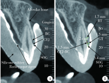
目的: 利用锥形束体层摄影术(cone-beam computed tomography,CBCT)分析牙周健康的汉族青年上颌前牙唇侧中央嵴顶上牙龈(supraosseous gingiva,SOG)的三维形态及相关解剖结构。方法: 选取25名牙周健康的汉族青年共计150颗上颌前牙纳入研究,受试者男性11名,女性14名,平均年龄(24.5±1.6)岁,佩戴含有显影剂的硅橡胶印模拍摄软组织间接显影CBCT。对影像资料进行三维重建并测量分析唇侧中央嵴顶上牙龈的形态,包括SOG高度、釉牙骨质界(cemento-enamel junction,CEJ)到骨嵴顶的距离、CEJ处牙龈厚度、骨嵴顶下2 mm牙槽骨厚度等。数据用SPSS 22.0软件进行统计分析,比较各牙位参数之间的差异,分析其相互之间的相关关系。结果: 上颌前牙唇侧中央SOG高度测量结果,中切牙为(3.54±0.67) mm、侧切牙为(3.48±0.81) mm、尖牙为(3.49±0.70) mm,各牙位SOG高度差异无统计学意义(P>0.05)。上颌前牙唇侧中央CEJ水平牙龈平均厚度测量结果,中切牙为(1.45±0.23) mm,侧切牙为(1.13±0.24) mm,尖牙为(1.14±0.22) mm,中切牙牙龈最厚,与侧切牙和尖牙相比差异均有统计学意义(P<0.05)。唇侧中央SOG与CEJ处牙龈厚度在所有牙位上均无明显相关性(P>0.05)。结论: 牙周健康的上颌前牙唇侧嵴顶上牙龈中,中切牙牙龈最厚,未发现上颌前牙区唇侧中央SOG高度与厚度存在相关性。
中图分类号:
- R783

| [1] |
Arora R, Narula S, Sharma R, et al. Supracrestal gingival tissue: Assessing relation with periodontal biotypes in a healthy periodon-tium [J]. Int J Periodontics Restorative Dent, 2013, 33(6):763-771.
doi: 10.11607/prd.1501 |
| [2] |
Perez JR, Smukler H, Nunn M E. Clinical evaluation of the supraosseous gingivae before and after crown lengthening [J]. J Periodontol, 2007, 78(6):1023-1030.
pmid: 17539715 |
| [3] |
Gargiulo AW, Wentz FM, Orban B. Dimensions and relations of the dentogingival junction in humans [J]. J Periodontol, 1961, 32(3):261-267.
doi: 10.1902/jop.1961.32.3.261 |
| [4] |
Kois JC. Altering gingival levels: The restorative connection part I: Biologic variables [J]. J Esthet Restor Dent, 1994, 6(1):3-7.
doi: 10.1111/j.1708-8240.1994.tb00825.x |
| [5] | Vacek JS, Gher ME, Assad DA, et al. The dimensions of the human dentogingival junction [J]. Int J Periodontics Restorative Dent, 1994, 14(2):154-165. |
| [6] |
Fischer KR, Grill E, Jockel-Schneider Y, et al. On the relationship between gingival biotypes and supracrestal gingival height, crown form and papilla height [J]. Clin Oral Implants Res, 2014, 25(8):894-898.
doi: 10.1111/clr.2014.25.issue-8 |
| [7] | 乐迪, 张豪, 胡文杰, 等. 牙周探诊法判断牙龈生物型的初步研究 [J]. 中华口腔医学杂志, 2012, 47(2):81-84. |
| [8] | 曹洁, 胡文杰, 张豪, 等. 基于锥形束计算机体层摄影术测量牙龈厚度 [J]. 北京大学学报(医学版), 2013, 45(1):135-139. |
| [9] | 张艳玲, 张豪, 胡文杰, 等. 120名汉族青年前段牙弓唇侧角化龈宽度的测量 [J]. 中华口腔医学杂志, 2010, 45(8):477-481. |
| [10] |
Zhang YL, Le D, Hu WJ, et al. Assessment of dynamic smile and gingival contour in young Chinese people [J]. Int Dent J, 2015, 65(4):182-187.
doi: 10.1111/idj.12174 |
| [11] |
Perez JR, Smukler H, Nunn ME. Clinical dimensions of the supraosseous gingivae in healthy periodontium [J]. J Periodontol, 2008, 79(12):2267-2272.
doi: 10.1902/jop.2008.080101 pmid: 19053916 |
| [12] |
Cao J, Hu WJ, Zhang H, et al. A novel technique for measurement of dentogingival tissue by cone beam computed tomography [J]. Oral Surg Oral Med Oral Pathol Oral Radiol, 2015, 119(2):e82-e87.
doi: 10.1016/j.oooo.2014.10.022 |
| [13] |
Alves PHM, Alves TCLP, Pegoraro TA, et al. Measurement pro-perties of gingival biotype evaluation methods [J]. Clin Implant Dent Relat Res, 2018, 20(3):280-284.
doi: 10.1111/cid.2018.20.issue-3 |
| [14] |
Hausmann E, Allen K, Clerehugh V. What alveolar crest level on a bite-wing radiograph represents bone loss? [J]. J Periodontol, 1991, 62(9):570-572.
pmid: 1941497 |
| [15] |
Ghassemian M, Nowzari H, Lajolo C, et al. The thickness of facial alveolar bone overlying healthy maxillary anterior teeth [J]. J Periodontol, 2012, 83(2):187-197.
doi: 10.1902/jop.2011.110172 pmid: 21692627 |
| [16] |
Nowzari H, Molayem S, Chiu CH, et al. Cone beam computed tomographic measurement of maxillary central incisors to determine prevalence of facial alveolar bone width ≥2 mm [J]. Clin Implant Dent Relat Res, 2012, 14(4):595-601.
doi: 10.1111/cid.2012.14.issue-4 |
| [17] |
Taylor R. Interpretation of the correlation coefficient: A basic review [J]. J Diagn Med Sonogr, 1990, 6(1):35-39.
doi: 10.1177/875647939000600106 |
| [18] | Kao RT, Fagan MC, Conte GJ. Thick vs thin gingival biotypes: A key determinant in treatment planning for dental implants [J]. J Calif Dent Assoc, 2008, 36(3):193-198. |
| [19] |
Müller HP, Könönen E. Variance components of gingival thickness [J]. J Periodontal Res, 2005, 40(3):239-244
pmid: 15853970 |
| [20] |
Fu JH, Yeh CY, Chan HL, et al. Tissue biotype and its relation to the underlying bone morphology [J]. J Periodontol, 2010, 81(4):569-574.
doi: 10.1902/jop.2009.090591 |
| [21] |
La Rocca AP, Alemany AS, Levi P Jr. Anterior maxillary and mandibular biotype: Relationship between gingival thickness and width with respect to underlying bone thickness [J]. Implant Dent, 2012, 21(6):507-515.
doi: 10.1097/ID.0b013e318271d487 |
| [22] | Frumkin N, Via S, Klinger A. Evaluation of the width of the alveolar bone in subjects with different gingival biotypes: A prospective cohort study using cone beam computed tomography [J]. Quintessence Int, 2017, 48(3):209-216. |
| [23] | Cook DR, Mealey BL, Verrett RG, et al. Relationship between clinical periodontal biotype and labial plate thickness: An in vivo study [J]. Int J Periodontics Restorative Dent, 2011, 31(4):345-354. |
| [24] |
Batista EL, Moreira CC, Batista FC, et al. Altered passive eruption diagnosis and treatment: A cone beam computed tomography-based reappraisal of the condition [J]. J Clin Periodontol, 2012, 39(11):1089-1096.
doi: 10.1111/j.1600-051X.2012.01940.x pmid: 22966787 |
| [1] | 白晓强, 袁芷若, 周永胜, 吕珑薇. 动态牵张促进人骨髓间充质干细胞三维培养的成骨分化[J]. 北京大学学报(医学版), 2026, 58(3): 641-649. |
| [2] | 温奥楠, 张晓会, 杨咏涛, 高梓翔, 李文博, 单珅瑶, 商相宜, 田淯文, 郭殊玮, 王艺蓁, 王勇, 赵一姣. 基于非刚性配准构建三维颜面微笑仿真序列数据的方法[J]. 北京大学学报(医学版), 2026, 58(1): 139-144. |
| [3] | 郑苗, 马欣蓉, 陈昊, 赵恒欣, 张宇, 谭建国, 李和平, 王霄. 大气压放电冷等离子体处理对人牙龈成纤维细胞生物学行为的影响[J]. 北京大学学报(医学版), 2026, 58(1): 60-67. |
| [4] | 邵梁, 马雯洁, 陈莹, 丁茜, 张磊. 上颌切牙前伸和正中咬合接触解剖特征的数字化测量与分析[J]. 北京大学学报(医学版), 2026, 58(1): 99-106. |
| [5] | 王翠萍, 陈哲, 程永静. 超微血流成像评估与膝骨关节炎临床症状的关联研究[J]. 北京大学学报(医学版), 2025, 57(6): 1096-1100. |
| [6] | 宋凤岐, 徐心雨, 刘筱菁, 李自力. 上颌骨前部和整体顺时针旋转改善骨性Ⅲ类牙颌面畸形患者鼻旁凹陷的对比[J]. 北京大学学报(医学版), 2025, 57(5): 980-988. |
| [7] | 肖宇嘉, 毛渤淳, 周彦恒. 姿势性微笑的三维形态学研究[J]. 北京大学学报(医学版), 2025, 57(5): 989-995. |
| [8] | 王泽远, 于栓宝, 郑浩轲, 陶金, 范雅峰, 张雪培. 基于临床特征和多参数MRI的前列腺癌盆腔淋巴结转移的术前预测模型[J]. 北京大学学报(医学版), 2025, 57(4): 684-691. |
| [9] | 宁圆, 张晓盈, 李雪, 李原, 何菁, 金月波. 干燥综合征并发乳腺淋巴瘤1例[J]. 北京大学学报(医学版), 2025, 57(4): 808-811. |
| [10] | 孙建军, 马千权, 尹晓亮, 杨辰龙, 张嘉, 陈素华, 吴超, 谢京城, 韩芸峰, 林国中, 司雨, 杨军, 邬海博, 赵强. 任意维度重建磁共振对骶管囊肿进行精准分型对于指导微创手术和康复的意义[J]. 北京大学学报(医学版), 2025, 57(2): 303-308. |
| [11] | 仇师禹, 练洋, 康一帆, 张雷, 蔡义望, 单小峰, 蔡志刚. 基于下颌骨数据库和全连接神经网络的三维检索模型辅助下的下颌骨个性化重建[J]. 北京大学学报(医学版), 2025, 57(2): 360-368. |
| [12] | 马丽娟, 腾雍辉, 王勇, 赵一姣, 张馨月, 秦庆钊, 尹东. 乳牙缺失数字化丝圈间隙保持器的三维有限元分析[J]. 北京大学学报(医学版), 2025, 57(2): 376-383. |
| [13] | 朱玉佳, 沈华, 温奥楠, 高梓翔, 秦庆钊, 单珅瑶, 李文博, 傅湘玲, 赵一姣, 王勇. 三维颌面对称参考平面智能构建的深度学习算法[J]. 北京大学学报(医学版), 2025, 57(1): 113-120. |
| [14] | 徐昕恺, 赵建江, 田素坤, 刘中宁, 赵晓一, 赵晓波, 江腾飞, 陈晓军, 马超, 孙玉春. 集成压缩气流系统扫描头辅助获取液体干扰表面三维数据精度评价[J]. 北京大学学报(医学版), 2025, 57(1): 121-127. |
| [15] | 吴灵, 方嘉琨, 刘筱菁, 李自力, 李阳, 王晓霞. 基于牙颌面畸形患者三维颅面特征相似性度量模型的建立及评估[J]. 北京大学学报(医学版), 2025, 57(1): 128-135. |
|
||
